Showing posts with label Traumatic Brain Injury. Show all posts

Monday, 19 September 2016

Preventing Depression Following Brain Injury

Depression is a common feature following traumatic brain injury (TBI). Post-TBI depression may be difficult to treat and evolve into a chronic depression syndrome.

A recent published study demonstrates that prophylactic SSRI antidepressant treatment may reduce the incidence of depression following TBI.

This study was conducted by investigators affiliated with Baylor College of Medicine and the Department of Psychiatry at the University of Iowa.

Ninety four subjects were recruited to this randomized control trial.

Active drug treatment was provided by the selective serotonin reuptake inhibitor sertraline. Subjects were dosed up to 100 mg per day of sertraline and followed for 24 weeks.

The primary findings of the study included the following:

  • Sertraline was well-tolerated with few adverse events in the study population
  • By 24 weeks approximately 25% of control subjects met criteria for depression while only about 7% of sertraline subjects met criteria for depression
  • The number of subjects needed to treat to prevent one person developing depression post-TBI was 5.9

The authors note in the discussion that sertraline and the SSRI class may be linked to:
"enhanced neuroplasticity in mood regulation areas, such as the hippocampus and prefrontal cortex"
The authors had previously noted an enhanced neuropsychological performance following TBI with the SSRI escitalopram. They were unable to replicate that finding in the current study.

This is an important study that deserves wider replication. If confirmed, SSRI primary prevention of depression following TBI may become a common practice.

Follow me on Twitter WRY999

Photo of monarch butterfly depositing egg on milkweed plant is from the author's files.

Jorge RE, Acion L, Burin DI, & Robinson RG (2016). Sertraline for Preventing Mood Disorders Following Traumatic Brain Injury: A Randomized Clinical Trial. JAMA psychiatry PMID: 27626622

Tuesday, 31 May 2016

The Promise of DTI (Imaging) in Neurology

Brain diffusion tensor imaging (DTI) is emerging as new important brain imaging tool in research.

Clinicial applications of DTI are lagging the research applications in development.

However, clinical applications are coming.

The Radiological Society of North America recently published a summary of potential application for DTI in Neurology on their website. You can access the website post HERE.

Three key areas were highlighted in this post including use of DTI for:
  • Aiding Identification of Prognosis in Mild TBI
  • Gender Effects on Sports-Related Head Injury
  • Brain Mapping Neurosurgery

Included at the website are two You Tube Videos. Here is the video related to using DTI as a prognostic aid in TBI.


Dr. ML Lipton featured in the You Tube video has a free full-text published a study of DTI in soccer heading in the manuscript cited below. Click on the PMID to get to the manuscript.

In this manuscript the authors were able to identify change in white matter brain microstructure related to prevalence of soccer heading independent of concussion.

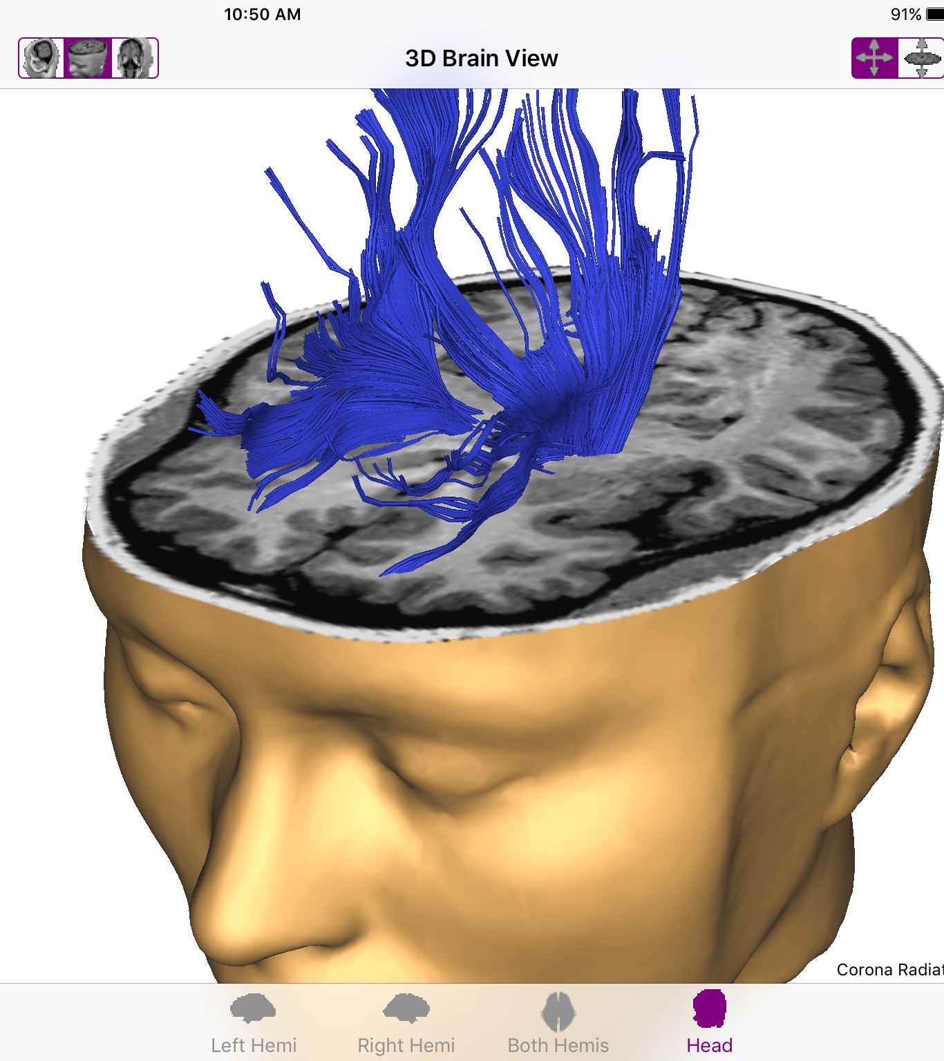
Image of white matter corona radiata is my screen shot from the iPad app Brain Tutor.

Follow me on Twitter WRY999

Lipton ML, Kim N, Zimmerman ME, Kim M, Stewart WF, Branch CA, & Lipton RB (2013). Soccer heading is associated with white matter microstructural and cognitive abnormalities. Radiology, 268 (3), 850-7 PMID: 23757503


Wednesday, 18 November 2015

Predictors of Poor Outcome After Traumatic Brain Injury

The outcome following traumatic brain injury (TBI) is often unpredictable and variable.

Two individuals with similar types of TBI can have quite different outcomes ranging from total disability to functional employment.

Torun Finnanger and colleagues from Norway and Australia recently reported on a study that examined a number of predictor variables on self-reported outcome following TBI.

In this study, 67 adolescents and adults with moderate to severe TBI completed baseline assessments and were followed for a period of 2 to 5 years. 

The key outcome variables in this study was self-reported executive, behavioral and emotional impairment at the follow-up time point. TBI subjects were compared on a variety of variables with a control group without injury.

Early assessment variables that were examined for correlation with outcome after TBI included:

  • Pre-trauma level of education
  • Brain injury severity rating: Glasgow Coma Score
  • Duration of post-traumatic amnesia
  • Brain imaging: presence of imaging markers for brain traumatic axonal injury on brain MRI with inclusion of diffusion tensor imaging (DTI) data
  • Neuropsychological assessment obtained three months after injury including: Motor function, information processing speed, attention, visual memory, verbal memory and executive function
  • Presence of the symptoms of depression using Beck Depression Inventory 3 and 12 months after injury

The key findings from this longitudinal predictor study included:

  • Self-reported impairment in executive function at 2-5 years follow up was linked to fewer years of education, early depression symptoms and traumatic axonal injury
  • Injury at a younger age predicted increased aggression and conduct disorder behavioral problems
  • Traumatic axonal injury and early depression symptoms predicted increased depression and anxiety symptoms at follow up.

The authors conclude their study supports inclusion of self-reported executive, emotional and behavioral assessments in the monitoring of TBI subjects.

Additionally, they note early MRI imaging assessment of axonal injury (in the first two weeks following TBI) and early assessment of depressive symptoms add information to standard neuropsychological assessment batteries.

They note accurately defining high-risk groups more poor outcome may allow targeting of more aggressive treatment strategies. 
"Furthermore, psychological and/or pharmaceutical interventions, with a focus on depressive symptomatology may be helpful in reducing the long-term problems experienced by persons sustaining a TBI.."

This study is filled with a ton of data. Readers with more interest in the study can find the free full-text manuscript by clicking on the PMID link in the citation below.

Follow the author on Twitter WRY999.

Image of brain white matter pathways is an iPad screen shot from the 3D Brain app from the authors files.

Finnanger TG, Olsen A, Skandsen T, Lydersen S, Vik A, Evensen KA, Catroppa C, HÃ¥berg AK, Andersson S, & Indredavik MS (2015). Life after Adolescent and Adult Moderate and Severe Traumatic Brain Injury: Self-Reported Executive, Emotional, and Behavioural Function 2-5 Years after Injury. Behavioural neurology, 2015 PMID: 26549936

Thursday, 1 October 2015

Does Brain Injury Increase Criminal Behavior?

Traumatic brain injury produces a variety of cognitive effects.

Some individuals will have behavioral disturbances including anger outbursts and interpersonal conflict.

A recent study examined the rates of first criminal conviction in a group of 7694 men and women hospitalized with traumatic brain injury (TBI).

The study found about a 50 to 75% increase in rates of post-TBI criminal convictions including violent convictions. This effect was noted using either general population controls or sibling controls.

Readers with more interest in this topic can find the abstract and link to the free full-text manuscript by clicking on the PMID link in the citation below.

Figure is an original chart abstracted from data in the manuscript.

Follow the author on Twitter @WRY999

Schofield PW, Malacova E, Preen DB, D'Este C, Tate R, Reekie J, Wand H, & Butler T (2015). Does Traumatic Brain Injury Lead to Criminality? A Whole-Population Retrospective Cohort Study Using Linked Data. PloS one, 10 (7) PMID: 26172545

Friday, 4 September 2015

Football and Brain Injury: Weekend Reading List

Here is a list of a few abstracts that caught my eye on the topic of football and brain injury risk.

I will be reviewing some of these in more detail for further discussion next week.

Clicking on the title will take you to the PubMed abstract. Selected abstracts have links to full text manuscripts.

This topic is an emerging area of interest and a easy summary of the state-of-the-art is "We have many unanswered questions".

Research is needed across many scientific domains including epidemiology, risk factors, detection, brain imaging and brain trauma biomarkers, genetics and treatment interventions.


Epidemiology of Sports-Related Concussion in NCAA Athletes From 2009-2010 to 2013-2014: Incidence, Recurrence, and Mechanisms.


Photo of milkweed flower,  a favorite of the monarch butterfly, is from the author's files.

Follow the author on Twitter: @WRY999

Tuesday, 17 March 2015

Physical Exercise Effects on Brain EEG

There are a variety of methods to study the effects on exercise on brain function. 

Brain imaging techniques such as fMRI provides a new tool to search for regional effects of acute and chronic exercise.

Another tool that has received less attention is the electroencephalogram or EEG. One EEG measure of brain function is the individual alpha peak frequency or iAPF.

The iAPF is positively correlated with arousal, attention and speed of information processing. Higher iAPF is linked to faster speed of information processing.

Boris Gutmann and colleagues from the German Sport University Cologne recently published a study of the effects on acute and chronic exercise on iAPF.

Here are the key elements in the design of their study:

  • Subjects: Ten males average age 22 who were regular participants in exercise but not highly trained endurance athletes
  • Exercise: Time1- iAPF before and after exhaustive exercise and before and after steady state. Time2-iAPF before and after exhaustive exercise and before and after steady state. Time 1 and Time 2 exercise sessions were separated by four weeks that included steady state aerobic exercise training

The key findings from the study were:

  • iAPF was increased after exhaustive exercise but not following steady state exercise
  • This effect occurred both at time 1 and time 2 suggesting it was independent of training

The authors note the arousal effect of exhaustive exercise may be "mediatede by changes in neural activity in the reticular-activating system". 

I found it interesting that the effects of iAPF was limited to exhaustive exercise. In the exhaustive exercise protocol, subjects rode stationary bikes controlled by the researchers for intensity. Subjects started at 30 watts increased by 5 watts every 30 seconds until subjects reached exhaustion. The steady state exercise protocol was exercise on the bicycle for 30 minutes at 65-75% of maximal heart rate.

There appears to be some increasing research interest in the value of shorter periods of exhaustive exercise like that seen in interval training protocols. Increasing evidence supports the value of brief high intensity periods of exercise in promoting health benefits of exercise.

Using EEG markers may be helpful in examining the potential value of exercise in a variety of brain disorders, i.e. traumatic brain injury, attention deficit disorder, mood and anxiety disorders. 

Readers with more interest in this study can access the free full-text manuscript by clicking on the PMID link below.

Photo of Captiva, FL sunset is from the author's files.

Follow the author on Twitter: @WRY999

Gutmann B, Mierau A, Hülsdünker T, Hildebrand C, Przyklenk A, Hollmann W, & Strüder HK (2015). Effects of physical exercise on individual resting state EEG alpha peak frequency. Neural plasticity, 2015 PMID: 25759762

Thursday, 22 September 2011

Is Lithium a Potential Aid in Traumatic Brain Injury?

Lithium carbonate serves as a primary treatment option in the treatment of mania and bipolar affective disorder.  An elemental metal, lithium has atomic number 3 in the periodic table of elements.

The mechanism of action for lithium carbonate in bipolar disorder is unclear.  Some of the proposed mechanisms for lithium in the central nervous system include:

  • alteration of the neurotransmitter glutamate (affected by other drugs linked to therapeutic effect in bipolar disorder, i.e. sodium valproate and lamotrigine)
  • alteration in gene expression
  • inactivation of the GSK-3B (glycogen synthase kinase) enzyme known to be involved in circadian clock regulation
  • interaction with the NO (nitrous oxide) signalling pathway
Inhibition of the GSK-3B enzyme has been shown to have potential beneficial effects in stimulating neuroplasticity as it is associated with enhanced expression of brain-derived neurotrophic factor (BDNF).  Fengshan Yu and colleagues at NIH and the University of Health Sciences have recently explored the effect of lithium on traumatic brain injury using a mouse model.

In their experiment, mice received doses of lithium chloride ranging form 1.0 to 5.0 mEq/kg dose of lithium or placebo following a controlled episode of brain trauma under anesthesia.  Doses were repeated daily for three days.

Brain injury response to lithium treatment was monitoring using neuropathological techniques as well as behavior and motor coordination tests.  The key results of the study include:
  • Lithium chloride at 1.5 to 3.0 mEq/kg reduced brain lesion volume compared to control
  • Lithium chloride reduced post-trauma related anxiety behavior during the outcome monitoring
  • Lithium chloride reduced breakdown of the blood-brain barrior
  • Short-term and long-term motor coordination was better in the lithium group

The authors note that their study suggests the neuroprotective effect of lithium administration following traumatic brain injury in the mouse model appears related to a GSK-3B mechanism.  The study timed lithium administration to 3 hours after the trauma providing a realistic model for a trial in human clinical scenarios.  They conclude "Our results that demonstrate its (lithium chloride) benefits in the mouse model pave the way for early clinical trials as potential treatment for TBI (traumatic brain injury) patients.

CT of traumatic brain injury showing cerebral contusion, cerebral hemmorhage, subdural hematoma and skull fracture from Wikipedia Creative Commons. Source: Rehman T, Ali R, Tawil I, Yonas H (2008). "Rapid progression of traumatic bifrontal contusions to transtentorial herniation: A case report". Cases journal 1 (1): 203. doi:10.1186/1757-1626-1-203. PMID 18831756. http://www.casesjournal.com/content/1/1/203


Yu F, Wang Z, Tchantchou F, Chiu CT, Zhang Y, & Chuang DM (2011). Lithium ameliorates neurodegeneration, suppresses neuroinflammation, and improves behavioral performance in a mouse model of traumatic brain injury. Journal of neurotrauma PMID: 21895523

Wednesday, 4 May 2011

Neuroethics: The Brain and Moral Beliefs

Jerome Grafman, Ph.D. presented the May 2011 Warren Frontiers in Neuroscience lecture “Brain Regions Supporting the Establishment of Human Beliefs” in Tulsa, Oklahoma.  I have typically summarized these lectures in a single Brain Posts blog posting.  But given the broad character of this presentation, I will break my summary into three parts based on the sections in the presentation: moral beliefs, religious beliefs and political beliefs.  Along with the lecture highlights, I will review relevant research manuscripts by Dr. Grafman and his research team.

A first step in studying concepts as abstract as morality is to define key components or factors within moral beliefs.  This is a common approach applicable to also religious and political beliefs.  Dr. Grafman prefers a more data driven approach to this task, rather than coming from a theoretical perspective.  The Moral Sentiment Task is a 98-item interview where subjects are given a scenario and asked to pick one of four responses that describes the feeling they would experience in that situation.  Using large population survey data, responses in this interview can be assigned to one of two sentiments:

Prosocial sentiments
  • embarrassment 
  • pity
  • guilt
Critical sentiments
  • anger
  • disgust
In a study with Jorge Moll and colleagues, Grafman examined a series of patients with frontotemporal dementia (FTD) using PET imaging and the Moral Sentiment Task.  Patients with FTD commonly exhibit impairment in social behaviors such as acting inappropriately (misjudge the social appropriateness of a behavior).  This socially inappropriate behavior commonly causes caregiver embarrassment and makes FTD often more difficult to manage than Alzheimer’s dementia.  Since the frontal lobe is felt to contribute to moral beliefs and judgment, a disease variably affecting the frontal lobe is likely to provide information about the effect of deficits in specific regions.

Patients with FTD were recruited for a study of the relationship between hypoactivation of specific brain regions with impairment in making accurate choices in the Moral Sentiment test.  As expected FTD, patients showed widespread decreased frontal lobe glucose utilization compared to controls.  In a correlational analysis, the following results were noted:
  • Decreased glucose utilization in the medial frontopolar cortex correlated with reduced prosocial sentiment
  • Decreased glucose utilization in the septum also correlated with impaired prosocial sentiment
  • Decreased glucose utilization in the dorsal medial prefrontal cortex and the amygdala correlated with reduced critical sentiment scores

In a review in the Annual Review of Neuroscience, Forbes and Grafman summarize the role of the prefrontal cortex in social cognition and moral judgment.  They note that there is significant overlap between processes involved in social cognition and moral judgment.  Social cognition has been described as the encoding, storage, retrieval and processing in the brain of information related to other members of the species. 

Elements of social cognition function can be implicit processes (occurring rapidly with little cognitive effort and outside conscious awareness) or explicit processes (deliberate, cognitively taxing and consciously accessible).  Implicit social cognition appears to have evolved earlier and is linked to the posterior cortex and subcortical regions.  Explicit social cognition evolved later and relies heavily on the prefrontal cortex (PFC). 

The prefrontal cortex appears to be a key area involved in making moral judgments.  Using fMRI brain PFC regions activate with processing a moral challenge.  Interestingly, the specific region of PFC involved in making a decision appears to be separate for making a decision on an impersonal moral dilemma (whether to flip a trolley track switch that will save five lives but lead to the death of one other person) or a personal moral dilemma (whether to smother your own crying baby if doing so would being detected by oppositional soldiers who will kill you and many other individuals).

Forbes and Grafman note in the conclusion of their review that “social interactions and judgments of humans have been based on evolutionary pressures and environmental and social contingencies”.  This evolution is continuing and they wonder if more impersonal world of advances such as Facebook and text messaging at a young age might drive the brain systems towards more “immediate results and gratification”.  Nevertheless, the emerging field of social neuroscience is likely to play a key role in better understanding neuroanatomical, genetic and environmental influences that effect our social development and the moral judgment we make.


Screen shot of prefrontal cortex from 3D Brain by Yates Photography.

In the next post, I will summarize research related to the brain and religious beliefs.  Dr. Grafman’s proposes an answer to the question “Is there a God spot in the brain?”

Moll J, Zahn R, de Oliveira-Souza R, Bramati IE, Krueger F, Tura B, Cavanagh AL, & Grafman J (2011). Impairment of prosocial sentiments is associated with frontopolar and septal damage in frontotemporal dementia. NeuroImage, 54 (2), 1735-42 PMID: 20728544


Forbes, C., & Grafman, J. (2010). The Role of the Human Prefrontal Cortex in Social Cognition and Moral Judgment Annual Review of Neuroscience, 33 (1), 299-324 DOI: 10.1146/annurev-neuro-060909-153230

Sunday, 24 April 2011

Recent Neuroscience Videos: TBI, Bipolar Disorder, Autism

This will be a little different Brain Post from the traditional research summaries I have posted in the past.  Instead this post will highlight some of the best videos I have recently run across related to neuroscience topics.  Most of these videos have a brief commercial prior to a video lasting 3 to 15 minutes.

Wall Street Journal: Nutrition Important to Healing Brain Injury-A brief discussion of issues related to providing the best nutrition in soldiers suffering a war-related brain injury.


Wall Street Journal: Shedding Light on Bipolar Disorder.  A six minute overview of bipolar disorder of recent intersest with the report that Catherine Zeta-Jones suffers from the disorder.


PBS NewsHour: The first in a series on autism from former anchor Robert McNeil.  This touching video shows McNeil's grandson who has autism.  It highlights the effects of autism on siblings and other family members.


Let me know if you find this format useful.  It will not be replacing the previous format but will become an occasional post to provide readers with some of the excellent video to better understand the disorders of clinical neuroscience.

Friday, 4 March 2011

Fluoxetine (Prozac) Boosts Motor Recovery After Stroke

Antidepressant drugs are being used for multiple non-depression indications including chronic pain, peripheral neuropathy, migraine prophylaxis, irritable bowel syndrome, hot flashes, premature ejaculation and insomnia (list not inclusive).   I had previously posted results of research looking at the effect of antidepressants after stroke on cognitive recovery.  This study from the University of Iowa College of Medicine, Department of Psychiatry found that escitalopram was linked to improved cognitive recovery following stroke compared to placebo.

Now on a related note, a study has been randomized placebo controlled study of fluoxetine has been published looking at recovery of motor function following acute ischemic stroke.   Here are the key elements of the design of this study:
  • Subjects:  113 patients with acute ischemic stroke ages 18-85 suffering hemiplegia or hemiparesis
  • Baseline motor deficit: Fugl-Meyer motor scale (FMMS) score of 55 or less
  • Intervention: Fluoxetine 20 mg daily for 3 months or placebo
  • Key outcome measure: FMMS score at day 90
The raters on the key outcome measure were blinded to the assigned treatment.  The findings from the study were impressive—mean (standard deviation) FMSS scores for the fluoxetine group was 54(28) compared to only 35.1 (22) in the placebo group.  As might be expected, rates of post-stroke depression were lower in the fluoxetine group (7% vs 29%).    Adverse events included nausea and diarrhea in the fluoxetine group—a typical selective serotonin reuptake inhibitor side effect.  One subject in each group died during the follow up period.  More patients in the fluoxetine group were more independent suggesting that motor recovery is an important contributor to global function after stroke.  The motor recovery effect appeared independent of lower depression rates in the fluoxetine group.

So why would a drug used typically for depression potentially have a role in post-stroke motor recovery?  The authors note animal studies have shown that drugs affecting brain neurotransmitters can modulate the rate and extent of recovery following brain injury.   Rats given fluoxetine after ischemic brain injury appear to have enhanced hippocampal neurogenesis.  These studies support a potential role for selective serotonin reuptake inhibitors like fluoxetine to have a neuroplasticity effect that may aid brain healing from a variety of insults.  This is not a magic bullet—it appears necessary to pair the drug treatment with active rehabilitation efforts.   But when used together, the combination appears to result in a better motor recovery after stroke.  I think we are likely to see more active research in this area and search for novel neuroprotective compounds that my provide better outcomes for stroke and other brain injury patients. 


Molecular model of fluoxetine molecule from Wikipedia Creative Commons, author Benjah-bmmj27.

Chollet F, Tardy J, Albucher JF, Thalamas C, Berard E, Lamy C, Bejot Y, Deltour S, Jaillard A, Niclot P, Guillon B, Moulin T, Marque P, Pariente J, Arnaud C, & Loubinoux I (2011). Fluoxetine for motor recovery after acute ischaemic stroke (FLAME): a randomised placebo-controlled trial. Lancet neurology, 10 (2), 123-30 PMID: 21216670

Wednesday, 26 January 2011

Kids and Brain Injury: TED Talk of Dr. Kim Gorgens


  • I tend to be a worrier and after having a child I had more things to worry about
  • I study brain injury in children
  • Leading emergency room causes for head injury in children: cycling, football, skateboard/skating
  • A 58 g force can occur in boxing,  a 103 g force can occur football
  • Average concussion 95 g force
  • Concussion: change in consciousness--being unconscious not required
  • Risk for second concussion greater than first
  • NFL study confirms 3 or more concussions increases risk for dementia
  • How do we guarantee the safety of our kids? Shows a picture of child surrounded by bubble wrap
  • I am trying to decide on allowing my child to play football--I haven't made a decision but we can all do three things

Study Up:learn all you can
Heads Up CDC--A education and prevention focused site that includes:
Concussion in Sports--resources for athletes and coaches
Information on Brain Injury for Physicians--includes link to Acute Concussion Evaluation form (ACE), a guideline for assessment of the athlete
Facebook Page for CDC Heads UP: Provides ongoing education and discussion forum on brain injury awareness
cokidswithbraininjury.com A resource for parents of children with brain injury--particularly in Colorado

Speak Up: talk to coaching staff and state legislature regarding traumatic brain injury

Suit UP: wear a helmet--bike helmets reduce brain impact by 50%

The comment section following this TED presentation notes this presentation was short on research documentation supporting Dr. Gorgens main points.  I agree, and I was unable to find any research published by her in a Pub Med search.  Nevertheless, this presentation highlights an important public health issue and provides some information resources for parents and clinicians. 

Friday, 14 January 2011

Brain Trauma: Getting the Best Care

An under-reported issue in the shooting of Representative Gabrielle Giffords is availability and access to high level trauma care. If you are going to survive a brain gun shot wound or other serious trauma it is crucial to quickly access specialized trauma hospitals and physicians to maximize your chance of survival.

Fortunately for Representative Giffords and the other survivors of the Tucson Arizona shootings, the incident occurred within only a few miles of a level I adult a trauma center. University Medical Center in Tucson has the highest rating for a trauma center. (Why the University of Arizona named their medical center University Medical Center is a mystery to me. By using a generic name they lost out on millions of dollars of free publicity with this incident.)

Trauma centers in the United States are categorized in five levels--I II III IV and V. According to Wikipedia, as of May 31, 2010 there were 203 level I trauma centers in the United States. The key distinctions for levels of rating are:

  • Level I: Highest level of surgical care for trauma, full range of physician surgical specialist available 24/7, significant annual number of trauma cases treated, surgical residency at site with significant educational and research program for trauma
  • Level II: Collaborates with a regional level I trauma center with 24 hour availability of essential physician specialties, less stringent annual trauma case volume, not required to participate in research or surgical residency training.
  • Level III: Full complement of surgical specialists not required but must provide emergency trauma care in collaboration with a level I or level II trauma center
  • Level IV and level V: Only require immediate 24 availability of trauma-trained nurse and fewer specialty physician availability requirements

A study published in the New England Journal of Medicine examined the role of trauma center care on mortality in the United States. Controlling for trauma case severity, they found mortality was reduced by 20 to 25% when trauma care was provided by a level I trauma center. This effect appeared to be particularly true for younger adults groups in the age range of Representative Giffords. This study was not specific to brain trauma or gun shot wounds to the brain. The effect of level I trauma care for this type of injury has not been extensively studied.

Academic medical centers such as the University of Arizona often benefit from 24 hour in-house surgical specialist residents.  These on-call residents can provide early assessment and management during the time it takes for surgical specialists to arrive at the hospital.

A Center for Disease Control website focuses on the availability of trauma centers in the U.S.   It highlights the geographic distribution of trauma centers in the United States and provides links to additional information. The map shows where you can get to a level I or level II within one hour via a helicopter transport.   The CDC site emphasizes the key in trauma care is: "Getting the Right Care, in the Right Place, at the Right Time".  Fortunately for Representative Giffords, she was only minutes away from a facility with the highest level of trauma care.

Photo of Palm Beach County Trauma Helicopter Courtesy of Wikipedia Creative Commons: Author Captain Mark Carr.

MacKenzie, E., Rivara, F., Jurkovich, G., Nathens, A., Frey, K., Egleston, B., Salkever, D., & Scharfstein, D. (2006). A National Evaluation of the Effect of Trauma-Center Care on Mortality New England Journal of Medicine, 354 (4), 366-378 DOI: 10.1056/NEJMsa052049