There is significant interest in activities that may boost academic achievement in the classroom. I previously posted on evidence that exercise prior to a learning task improved reading comprehension scores. You can access that post by clicking HERE. Now a study has compared two types of activities after a memorization task in male students. In this study, 60 male students completed a learning task and then were randomized into one of three activities for one hour. The three activities were playing a violent video game, a period of running or a control period of conversation. After the activity period, subjects completed a memory test. Subjects had salivary cortisol levels examined before and after the learning task at at the time of memory testing. All subjects had a increase in salivary cortisol levels after the learning phase, but only the running group demonstrated a continued rise in cortisol after the activity phase. The subjects participating in a running activity performed better on the memory retention test than those in the violent video game and the conversation control groups. There was no correlation between salivary cortisol levels and memory retention. The authors conclude that their finding has implications for scheduling exercise during the school day for children. They recommend physical exercise following intense learning cycles to promote improved learning efficiency. Interestingly, in the United States many schools are cutting back on physical education activities due to budget constraints. Such cutbacks may contribute to declining performance on testing metrics. Readers with more interest in this topic can access the free full text manuscript by clicking on the DOI link in the citation below. Photo of monarch butterfly is from the author's files. Follow me on Twitter at WRY999 Kindermann, H., Javor, A., & Reuter, M. (2016). Playing counter-strike versus running: The impact of leisure time activities and cortisol on intermediate-term memory in male students Cognitive Systems Research, 40, 1-7 DOI: 10.1016/j.cogsys.2016.01.002
Child brain development benefits from a positive parenting style and environment. The mechanism for this positive effect is unclear but moderation of the stress response in the growing child is an area of research interest. Haroon Sheikh and colleagues from the University of Ontario in Canada recently published results on a study of parenting and brain development in children. In their study, a cohort of 46 six year old girls underwent brain imaging using a technique known as diffusion tensor imaging or DTI. DTI provides a measure of brain white matter integrity. This study is informative because all the girls participated in an earlier study of stress reactivity at three years of age. High stress reactivity as measured by serum cortisol response is known to be linked to vulnerability to mood and anxiety disorders. The key elements in the design of this study including the following:
Subjects: 45 six year old girls from a larger ongoing longitudinal study of children
Stress response status: At three years of age participants underwent a two phase study of stress response. A baseline salivary cortisol assay was collected. A second cortisol level was obtained during a stressful task. Subjects were grouped in four categories based on levels of cortisol.
Parenting assessment: Parents and child participated in a play task. Parents were rated on a measure of parental negative and positive affect.
MRI scanning: A 3 Telsa brain imaging scan was completed on average two and one-half years following the baseline cortisol and parenting assessment
The main findings from the study included the following:
High stress reactivity at baseline was linked to lower white matter integrity in prefrontal and basal brain regions (left thalamus, right anterior cingulate cortex and right superior frontal gyrus)
Positive parental affectivity reduced the brain white matter effects of stress(cortisol) response in the right anterior cingulate cortex and right superior frontal gyrus
Children with high stress responses at baseline but a positive parental affect environment showed brain integrity findings similar to low stress reactivity children
This is an important study because it suggests an interaction between parental environment and adverse effects of a high stress response in three year old girls. Genetic factors likely contribute to level of stress response in three year old girls. A positive parental style appears to reduce or eliminate adverse effects of high stress reactivity on critical white matter brain development. The implications of the study are important. High-risk children for mood and anxiety disorders may benefit from early identification and parental training to reduce risk for later psychological morbidity. Readers with more interest in this study can access the free full-text manuscript by clicking on the PMID link below. Photo of hawk in flight is from the author's files. Follow the author on Twitter @WRY999 Sheikh HI, Joanisse MF, Mackrell SM, Kryski KR, Smith HJ, Singh SM, & Hayden EP (2014). Links between white matter microstructure and cortisol reactivity to stress in early childhood: evidence for moderation by parenting. NeuroImage. Clinical, 6, 77-85 PMID: 25379418
One of the myths surrounding ketogenic diets comes from misunderstanding the role of cortisol — the "stress hormone".
In a previous post, we addressed one of the arguments behind this myth: the idea that to activate gluconeogenesis (to make glucose out of protein), extra cortisol must be recruited. That is just factually incorrect, as we showed in the post.
The other argument, which we address here, is more complex.
Like the previous cortisol myth, it involves a faulty chain of reasoning. Here are the steps:
Ketogenic diets may raise certain measures of cortisol.
Chronically elevated cortisol is correlated with metabolic sydrome, and therefore higher cortisol measures may indicate the onset of metabolic syndrome.
Therefore, ketogenic diets could cause metabolic syndrome.
Metabolic syndrome is a terrible and prevalent problem today. It is that cluster of symptoms most strongly identified with diabetes — excess abdominal fat, high blood sugar, and a particular cholesterol profile — but also correlated with other life-threatening conditions such as heart disease and cancer.
In this post, we're going to explain some of the specifics of cortisol metabolism. We'll show how this argument is vague, and how clarifying it leads to the opposite conclusion. The confusion may all stem from misunderstanding one important fact: different measures of cortisol are not equivalent.
First, though, there is an important reason why the argument doesn't make sense.
We already know that a ketogenic diet effectively treats metabolic syndrome. As we will describe below, it turns out that certain cortisol patterns are strongly linked to metabolic syndrome, and might even be a cause of metabolic syndrome. If the cortisol pattern that develops in response to a ketogenic diet were the kind that was associated with metabolic syndrome, then we would expect people on ketogenic diets to show signs of abdominal fat gain, rising blood sugar, and a worsening cholesterol profile, but we see the opposite. This by itself makes it highly unlikely that ketogenic diets raise cortisol in a harmful way.
In other words, because cortisol regulation is so deeply connected to metabolic syndrome, the fact that ketogenic diets reverse symptoms of metabolic syndrome is itself strong evidence that theyimprovecortisol metabolism.
In Brief
There are many different measures of cortisol, because researchers have identified many different processes in cortisol metabolism.
Increases in some of those measurements are consistently linked to metabolic syndrome, and others are not.
Some researchers believe that cortisol dysregulation is a key underlying factor in metabolic syndrome.
The cornerstone of this connection may be the activity of an enzyme, 11β-HSD1. It converts from the inactive form cortisone to the active cortisol.
In metabolic syndrome, 11β-HSD1 is underactive in liver tissue and overactive in fat tissue. This results in a high rate of cortisol clearance, and low rate of regeneration.
These symptoms of cortisol dysregulation associated with metabolic syndrome were found to be reversed by a keto diet in a study that made the necessary measurements.
Does a ketogenic diet raise cortisol?
Boston Children's Hospital graphic (with our markup in black). Click for the original.
In a widely-cited study [1], from the Harvard-affiliated Boston Children's Hospital, published in the Journal of the American Medical Association, three different diets were tested: a low-fat diet, a low-carb diet, and a low-glycemic-index diet. The study showed that the different diets had substantially different metabolic effects, with the low-carbohydrate diet having the best results. To our surprise, the researchers then recommended the low-glycemic-index diet instead. As they explained in the accompanying press release:
“The very low-carbohydrate diet produced the greatest improvements in metabolism, but with an important caveat: This diet increased participants' cortisol levels, which can lead to insulin resistance and cardiovascular disease.”
The Boston Children's Hospital then went on to produce a graphic advising patients to follow the low-glycemic-index diet, and giving this as the primary reason not to choose the low-carb diet. Here is that graphic, which we've marked (in black) to show our disagreement. (Click for the full version without our markup.)
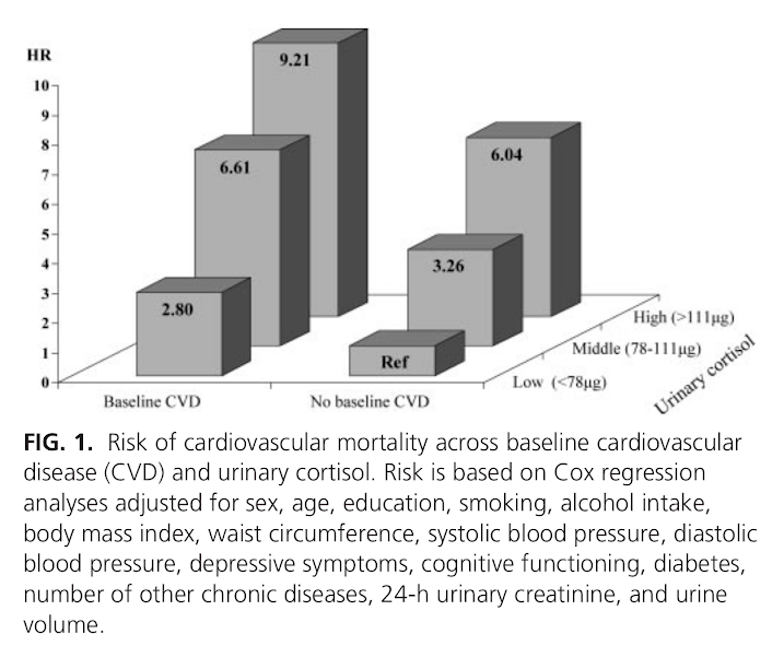
The cortisol levels are an understandable concern, because high urinary cortisol has been epidemiologically associated with a greatly increased risk of death from heart attacks [2].
However, because a ketogenic diet effectively treats metabolic syndrome, we should expect that it also reduces those specific cortisol patterns that are associated with metabolic syndrome (and therefore heart disease). As we show below, this has, in fact, been found.
How is cortisol associated with metabolic syndrome?
Cortisol can be measured in fluids, such as urine, saliva, or blood. Within those fluids, the amount of free cortisol can be measured, but so can cortisone, the inactive form, or the metabolites that are the result of enzyme action, and the ratios of any of these to the others can be measured (see Figure 1). Moreover, these measurements have a diurnal rhythm, being higher and lower at different times of the day.
The enzyme 11β-hydroxysteroid dehydrogenase (11β-HSD) can convert back and forth between cortisol and cortisone. 11β-HSD1—a subtype of 11β-HSD—converts cortisone to cortisol. When inactive cortisone is converted to the active cortisol, it is called regeneration. The other enzymes in the illustration break cortisone or cortisol down into metabolites. That process is called clearance. It turns out that measurements of these enzyme are important for evaluating cortisol metabolism.
The cortisol profile that has been associated with metabolic syndrome includes the following characteristics:
high 11β-HSD1 expression in fat cells, and low 11β-HSD1 expression in the liver [6], [7], which determines when and where cortisol is regenerated.
Similarly to the way total cholesterol measurement is correlated with heart disease, but only because it is roughly correlated with more informative cholesterol measurements, 24-hour urinary cortisol may be a proxy for production or clearance, but a poor one [3], [4], [7].
Cortisol levels are affected by production, but they are also affected by regeneration and clearance. In other words, if regeneration were increased, or clearance decreased, levels could go up even if production stayed the same or went down. (We previously discussed a similar situation with blood glucose and faulty inference about glucose production rates.) This means that levels can look similar, even when cortisol metabolism is very different.
Implication for those following the “adrenal fatigue” hypothesis: if you measure your cortisol, and it is high, you can't conclude that your adrenal glands are working correspondingly hard. It could be due to increased regeneration and reduced clearance by enzyme activity. Higher cortisol could actually mean the adrenals are working less!
In obesity, it appears that production goes up to compensate for high clearance and impaired regeneration, although sometimes not enough to compensate; blood cortisol is sometimes actually lower in obese subjects [8].
How does a ketogenic diet affect the relevant cortisol measures?
In [9], investigators put obese men on either a high-fat/low-carb (fat 66%, carb 4%) or a moderate-fat/moderate-carb (fat 35%, carb 35%) diet ad libitum (eating as much as they wanted). Note that both diets had the same protein percent, and both were lower carb than a standard American diet, but only the high-fat/low-carb diet was at ketogenically low levels.
For the high-fat/low-carb group, “the metabolic syndrome pattern” was reversed: blood cortisol went up, clearance went down, and regeneration went up. This was apparently due to an increase of 11β-HSD1 activity in liver tissue.
(Activity of 11β-HSD1 did not go down in fat tissue of those subjects, but the authors point out that the activity in fat tissue tends to go down when more fat is eaten, and the high-fat/low-carb group weren't actually eating more fat in absolute terms than at baseline, only lower carb.)
This reversal didn't happen in the moderate-fat/moderate-carb group, even though they lost a similar amount of weight.
So the ketogenic diet actually improved the cortisol profile of the participants, making it less like the cortisol profile seen in metabolic syndrome.
Summary
There is some reason to believe that cortisol dysregulation is a key underlying factor in metabolic syndrome [10], [11]. The dysregulation has a particular pattern that seems to be caused by a tissue-specific expression of the enzyme 11β-HSD1.
There is a belief among some researchers that ketogenic diets worsen cortisol metabolism (which could lead to metabolic syndrome and heart disease), but an examination of the specific pattern of cortisol metabolism related to metabolic sydrome shows the opposite.
This is what should have been expected in the first place, since ketogenic diets have already been shown to improve insulin sensitivity (the defining symptom of metabolic syndrome) in repeated randomized controlled trials.
One mechanism by which keto diet improves metabolic syndrome may be its beneficial effect on cortisol metabolism.
Ebbeling CB, Swain JF, Feldman HA, Wong WW, Hachey DL, Garcia-Lago E, Ludwig DS.
JAMA. 2012 Jun 27;307(24):2627-34. doi: 10.1001/jama.2012.6607.
(Emphases ours)
"Participants Overweight and obese young adults (n=21).
"Interventions After achieving 10 to 15% weight loss on a run-in diet, participants consumed low-fat (LF; 60% of energy from carbohydrate, 20% fat, 20% protein; high glycemic load), low-glycemic index (LGI; 40%-40%-20%; moderate glycemic load), and very-low-carbohydrate (VLC; 10%-60%-30%; low glycemic load) diets in random order, each for 4 weeks.
…
"Hormones and Components of the Metabolic Syndrome (Table 3)
"Serum leptin was highest with the LF diet (14.9 [12.1 to 18.4] ng/mL), intermediate with the LGI diet (12.7 [10.3 to 15.6] ng/mL) and lowest with the VLC diet (11.2 [9.1 to 13.8] ng/mL; P=0.0006). Cortisol excretion measured with a 24-hour urine collection (LF: 50 [41 to 60] μg/d; LGI: 60 [49 to 73] μg/d; VLC: 71 [58 to 86] μg/d; P=0.005) and serum TSH (LF: 1.27 [1.01 to 1.60] μIU/mL; LGI: 1.22 [0.97 to 1.54] μIU/mL; VLC: 1.11 [0.88 to 1.40] μIU/mL; P=0.04) also differed in a linear fashion by glycemic load. Serum T3 was lower with the VLC diet compared to the other two diets (LF: 121 [108 to 135] ng/dL; LGI: 123 [110 to 137] ng/dL; VLC: 108 [96 to 120] ng/dL; P=0.006).
"Regarding components of the metabolic syndrome, indexes of peripheral (P=0.02) and hepatic (P=0.03) insulin sensitivity were lowest with the LF diet. Serum HDL-cholesterol (LF: 40 [35 to 45] mg/dL; LGI: 45 [41 to 50] mg/dL; VLC: 48 [44 to 53] mg/dL; P<0.0001), triglycerides (LF: 107 [87 to 131] mg/dL; LGI: 87 [71 to 106] mg/dL; VLC: 66 [54 to 81] mg/dL; P<0.0001), and PAI-1 (LF: 1.39 [0.94 to 2.05] ng/mL; LGI: 1.15 [0.78 to 1.71] ng/mL; VLC: 1.01 [0.68 to 1.49] ng/mL; P for trend=0.04) were most favorable with the VLC diet and least favorable with the LF diet.
…
"Although the very low-carbohydrate diet produced the greatest improvements in most metabolic syndrome components examined here, we identified two potentially deleterious effects of this diet. Twenty-four hour urinary cortisol excretion, a hormonal measure of stress, was highest with the very low-carbohydrate diet. Consistent with this finding, Stimson et al reported increased whole-body regeneration of cortisol by 11β-HSD1 and reduced inactivation of cortisol by 5α-and 5β-reductases over 4 weeks on a VLC vs. a moderate-carbohydrate diet. Higher cortisol levels may promote adiposity, insulin resistance, and cardiovascular disease, as observed in epidemiological studies."
Comment: It is ironic that the authors bring up Stimson et al. as an example of a study that corroborates their findings. This is the very study[9]that, in our opinion, exonerates the VLC diet with respect to cortisol.
"Context: The stress hormone cortisol has been linked with unfavorable cardiovascular risk factors, but longitudinal studies examining whether high levels of cortisol predict cardiovascular mortality are largely absent.
Objective: The aim of this study was to examine whether urinary cortisol levels predict all-cause and cardiovascular mortality over 6 yr of follow-up in a general population of older persons.
Design and Setting: Participants were part of the InCHIANTI study, a prospective cohort study in the older general population with 6 yr of follow-up.
Participants: We studied 861 participants aged 65 yr and older.
Main Outcome Measure: Twenty-four-hour urinary cortisol levels were assessed at baseline. In the following 6 yr, all-cause and cardiovascular mortality was ascertained from death certificates. Cardiovascular mortality included deaths due to ischemic heart disease and cerebrovascular disease.
Results: During a mean follow-up of 5.7 (sd = 1.2) yr, 183 persons died, of whom 41 died from cardiovascular disease. After adjustment for sociodemographics, health indicators, and baseline cardiovascular disease, urinary cortisol did not increase the risk of noncardiovascular mortality, but it did increase cardiovascular mortality risk. Persons in the highest tertile of urinary cortisol had a five times increased risk of dying of cardiovascular disease (hazard ratio = 5.00; 95% confidence interval = 2.02–12.37). This effect was found to be consistent across persons with and without cardiovascular disease at baseline (p interaction = 0.78).
Conclusions: High cortisol levels strongly predict cardiovascular death among persons both with and without preexisting cardiovascular disease. The specific link with cardiovascular mortality, and not other causes of mortality, suggests that high cortisol levels might be particularly damaging to the cardiovascular system."
Jonathan Q. Purnell, Steven E. Kahn, Mary H. Samuels, David Brandon, D. Lynn Loriaux, and John D. Brunzell
Am J Physiol Endocrinol Metab. 2009 February; 296(2): E351–E357.
"Controversy exists as to whether endogenous cortisol production is associated with visceral obesity and insulin resistance in humans. We therefore quantified cortisol production and clearance rates, abdominal fat depots, insulin sensitivity, and adipocyte gene expression in a cohort of 24 men. To test whether the relationships found are a consequence rather than a cause of obesity, eight men from this larger group were studied before and after weight loss. Daily cortisol production rates (CPR), free cortisol levels (FC), and metabolic clearance rates (MCR) were measured by stable isotope methodology and 24-h sampling; intra-abdominal fat (IAF) and subcutaneous fat (SQF) by computed tomography; insulin sensitivity (SI) by frequently sampled intravenous glucose tolerance test; and adipocyte 11β-hydroxysteroid dehydrogenase-1 (11β-HSD-1) gene expression by quantitative RT-PCR from subcutaneous biopsies. Increased CPR and FC correlated with increased IAF, but not SQF, and with decreased SI. Increased 11β-HSD-1 gene expression correlated with both IAF and SQF and with decreased SI. With weight loss, CPR, FC, and MCR did not change compared with baseline; however, with greater loss in body fat than lean mass during weight loss, both CPR and FC increased proportionally to final fat mass and IAF and 11β-HSD-1 decreased compared with baseline. These data support a model in which increased hypothalamic-pituitary-adrenal activity in men promotes selective visceral fat accumulation and insulin resistance and may promote weight regain after diet-induced weight loss, whereas 11β-HSD-1 gene expression in SQF is a consequence rather than cause of adiposity.
"Previous studies have shown that compared with women, men have increased CPR (29), cortisol levels (29, 44), and visceral adiposity (9, 13). Given that hypercortisolemia can induce central obesity in disease states such as Cushing's syndrome, elevated endogenous cortisol secretion has been considered a potential mechanism that contributes to the expression of visceral adiposity in humans. However, of four previous reports that used 24-h urinary excretion rates of cortisol as a surrogate for cortisol production, only one found significant relationships between urinary secretion of total glucocorticoids, truncal fat, and insulin sensitivity in men and women (39), while three other studies in men have failed to show associations between urinary glucocorticoid secretion and either WHR (16, 26) or visceral fat (48). These studies, however, did not measure cortisol production directly, did not include blood FC, and did not test for differences in circadian variations of blood levels of cortisol, and in only one study was visceral fat specifically measured.
[...]
"In summary, we found in men that increased CPR and circulating FC are associated with accumulation of IAF, but not SQF, and with insulin resistance and impaired islet β-cell compensation (DI)."
Diabetologia. 2007 May;50(5):1024-32. Epub 2007 Mar 17.
"AIMS/HYPOTHESIS: The regulation of cortisol metabolism in vivo is not well understood. We evaluated the relationship between cortisol metabolism and insulin sensitivity, adjusting for total and regional fat content and for non-alcoholic fatty liver disease.
"MATERIALS AND METHODS: "Twenty-nine middle-aged healthy men with a wide range of BMI were recruited. We measured fat content by dual-energy X-ray absorptiometry and magnetic resonance imaging (MRI), liver fat by ultrasound and MRI, the hypothalamic-pituitary-adrenal axis by adrenal response to ACTH(1-24), unconjugated urinary cortisol excretion, corticosteroid-binding globulin, and cortisol clearance by MS. We assessed insulin sensitivity by hyperinsulinaemic-euglycaemic clamp and by OGTT.
"RESULTS: "Cortisol clearance was strongly inversely correlated with insulin sensitivity (M value) (r = -0.61, p = 0.002). Cortisol clearance was increased in people with fatty liver compared with those without (mean+/-SD: 243 +/- 10 vs 158 +/- 36 ml/min; p = 0.014). Multiple regression modelling showed that the relationship between cortisol clearance and insulin sensitivity was independent of body fat. The relationship between fatty liver and insulin sensitivity was significantly influenced by body fat and cortisol clearance.
"CONCLUSIONS/INTERPRETATION: "Cortisol clearance is strongly associated with insulin sensitivity, independently of the amount of body fat. The relationship between fatty liver and insulin sensitivity is mediated in part by both fatness and cortisol clearance."
[...]
"Since we showed no strong associations between measures of insulin sensitivity and 09.00 h cortisol levels, ACTH-stimulated cortisol concentrations, and unconjugated urinary cortisol excretion, these findings suggest that the relationship between these other aspects of cortisol metabolism and insulin sensitivity is relatively weak."
"The present study was designed to examine the hypothesis that hypothalamic-pituitary-adrenal axis activity as measured by 24-h cortisol production rate (CPR) and plasma levels of free cortisol is linked to increased body fat in adults, and that increased cortisol levels with aging results from increased CPR. Fifty-four healthy men and women volunteers with a wide range of body mass indexes and ages underwent measurement of CPR by isotope dilution measured by gas chromatography-mass spectroscopy, cortisol-binding globulin, and free cortisol in pooled 24-h plasma, body composition, and leptin. Cortisol clearance rates were determined from the 10-h disappearance curves of hydrocortisone after steady-state infusion in a separate group of lean and obese subjects with adrenal insufficiency. Although CPR significantly increased with increasing body mass index and percentage body fat, free cortisol levels remained independent of body composition and leptin levels due to increased cortisol clearance rates. CPR and free cortisol levels were, however, significantly higher in men than women. In addition, 24-h plasma free cortisol levels were increased with age in association with increased CPR, independent of body size. This increase in hypothalamic-pituitary-adrenal axis activity may play a role in the alterations in body composition and central fat distribution in men vs. women and with aging."
Arq Bras Endocrinol Metabol. 2007 Nov;51(8):1397-403.
"Human studies
"The bulk of evidences points both to an overexpression and an increased activity of 11bHSD1 in subcutaneous (SAT) and visceral adipose tissue (VAT) of obese subjects, although biopsies of the omentum were conducted in but a few studies. Several groups have shown higher 11bHSD1 mRNA expression in obese compared to non-obese subjects (29-32), although not all studies agree (33). Direct in vivo measurements using microdialysis in SAT also suggest an increase in the conversion rate of cortisone to cortisol (34). Moreover, 11bHSD1 mRNA expression positively correlates with obesity (body mass index and abdominal circumference), body composition, insulin resistance (30-32), resistins and other cytokines, as TNFa, IL-6, and leptin (35).
"The whole body 11bHSD1 activity reflects mainly hepatic expression. Initial studies that relied on measurements of cortisol-to-cortisone metabolites in urine (23,36) should be taken with caution as indicative of 11bHSD1 activity, because several other cortisol and cortisone metabolizing enzymes are deregulated in obesity (36). Of greater importance is the finding of reduced hepatic 11bHSD1 activity measured by the conversion of orally administered cortisone to cortisol (23,37). Thus, 11bHSD1 upregulation in obesity seems not to be a generalized process. In both the whole body and the splanchnic circulation there are no differences between obese and lean subjects regarding cortisol regeneration rates (as measured by [2H4]-cortisol tracer), presumably because an upregulation in adipose tissue is counterbalanced by a downregulation in the liver (15).
"Polymorphisms in the 11bHSD1 gene were identified in an attempt to clarify the basis for the increased activity of adipose tissue 11bHSD1 in obesity. In two populations, polymorphisms were associated with an increased risk of diabetes and hypertension, but not obesity (38,39). A polymorphism was also found that predicts lower 11bHSD1 expression and protection against diabetes (40)."
Wake DJ, Rask E, Livingstone DE, Söderberg S, Olsson T, Walker BR.
J Clin Endocrinol Metab. 2003 Aug;88(8):3983-8.
(emphasis ours)
"In idiopathic obesity circulating cortisol levels are not elevated, but high intraadipose cortisol concentrations have been implicated. 11beta-Hydroxysteroid dehydrogenase type 1 (11HSD1) catalyzes the conversion of inactive cortisone to active cortisol, thus amplifying glucocorticoid receptor (GR) activation. In cohorts of men and women, we have shown increased ex vivo 11HSD1 activity in sc adipose tissue associated with in vivo obesity and insulin resistance. Using these biopsies, we have now validated this observation by measuring 11HSD1 and GR mRNA and examined the impact on intraadipose cortisol concentrations, putative glucocorticoid regulated adipose target gene expression (angiotensinogen and leptin), and systemic measurements of cortisol metabolism. From aliquots of sc adipose biopsies from 16 men and 16 women we extracted RNA for real-time PCR and steroids for immunoassays. Adipose 11HSD1 mRNA was closely related to 11HSD1 activity [standardized beta coefficient (SBC) = 0.58; P < 0.01], and both were positively correlated with parameters of obesity (e.g. for BMI, SBC = 0.48; P < 0.05 for activity, and SBC = 0.63; P < 0.01 for mRNA) and insulin sensitivity (log fasting plasma insulin; SBC = 0.44; P < 0.05 for activity, and SBC = 0.33; P = 0.09 for mRNA), but neither correlated with urinary cortisol/cortisone metabolite ratios. Adipose GR-alpha and angiotensinogen mRNA levels were not associated with obesity or insulin resistance, but leptin mRNA was positively related to 11HSD1 activity (SBC = 0.59; P < 0.05) and tended to be associated with parameters of obesity (BMI: SBC = 0.40; P = 0.09), fasting insulin (SBC = 0.65; P < 0.05), and 11HSD1 mRNA (SBC = 0.40; P = 0.15). Intraadipose cortisol (142 +/- 30 nmol/kg) was not related to 11HSD1 activity or expression, but was positively correlated with plasma cortisol. These data confirm that idiopathic obesity is associated with transcriptional up-regulation of 11HSD1 in adipose, which is not detected by conventional in vivo measurements of urinary cortisol metabolites and is not accompanied by dysregulation of GR. Although this may drive a compensatory increase in leptin synthesis, whether it has an adverse effect on intraadipose cortisol concentrations and GR-dependent gene regulation remains to be established."
Proceedings of the Nutrition Society / Volume 66 / Issue 01 / February 2007, pp 1-8
(emphasis ours)
"The parallels between the clinical features of Cushing’s syndrome and the features of the metabolic syndrome (visceral obesity, hyperglycaemia, hypertension) led to the hypothesis that obesity is associated with glucocorticoid excess (Bjorntorp, 1991). In several monogenic rodent models obesity is accompanied by elevated circulating glucocorticoid levels, and the obesity is prevented by adrenalectomy. Hyperactivity of the HPA axis was thought to reflect chronic stress. However, although there is some evidence for greater stress responsiveness of the HPA axis in obesity (Rosmond et al . 1998; Epel et al . 2000), stress does not appear to explain HPA axis activation in metabolic syndrome (Brunner et al . 2002). Most importantly, in human obesity it appears that cortisol secretion (Marin et al . 1992; Pasquali et al . 1993) is increased to match elevated metabolic clearance (Strain et al . 1982; Andrew et al . 1998; Lottenberg et al . 1998), and does not result in increased plasma cortisol concentrations. Indeed, plasma cortisol concentrations are generally lower amongst obese subjects (Ljung et al . 1996; Walker et al . 2000; Reynolds et al . 2003), i.e. inverse to the effects on the HPA axis seen during starvation (see earlier; p. 2)."
"CONTEXT: Dietary macronutrient composition influences cardiometabolic health independently of obesity. Both dietary fat and insulin alter glucocorticoid metabolism in rodents and, acutely, in humans. However, whether longer-term differences in dietary macronutrients affect cortisol metabolism in humans and contribute to the tissue-specific dysregulation of cortisol metabolism in obesity is unknown.
OBJECTIVE: The objective of the study was to test the effects of dietary macronutrients on cortisol metabolism in obese men.
DESIGN: The study consisted of two randomized, crossover studies.
SETTING: The study was conducted at a human nutrition unit.
PARTICIPANTS: Participants included healthy obese men.
INTERVENTIONS, OUTCOME MEASURES, AND RESULTS: Seventeen obese men received 4 wk ad libitum high fat-low carbohydrate (HF-LC) (66% fat, 4% carbohydrate) vs. moderate fat-moderate carbohydrate (MF-MC) diets (35% fat, 35% carbohydrate). Six obese men participated in a similar study with isocaloric feeding. Both HF-LC and MF-MC diets induced weight loss. During 9,11,12,12-[(2)H](4)-cortisol infusion, HF-LC but not MF-MC increased 11beta-hydroxysteroid dehydrogenase type 1 (11beta-HSD1) activity (rates of appearance of cortisol and 9,12,12-[(2)H](3)-cortisol) and reduced urinary excretion of 5alpha- and 5beta-reduced [(2)H](4)-cortisol metabolites and [(2)H](4)-cortisol clearance. HF-LC also reduced 24-h urinary 5alpha- and 5beta-reduced endogenous cortisol metabolites but did not alter plasma cortisol or diurnal salivary cortisol rhythm. In sc abdominal adipose tissue, 11beta-HSD1 mRNA and activity were unaffected by diet.
CONCLUSIONS: A low-carbohydrate diet alters cortisol metabolism independently of weight loss. In obese men, this enhances cortisol regeneration by 11beta-HSD1 and reduces cortisol inactivation by A-ring reductases in liver without affecting sc adipose 11beta-HSD1. Alterations in cortisol metabolism may be a consequence of macronutrient dietary content and may mediate effects of diet on metabolic health."
"Recent evidence strongly argues for a pathogenic role of glucocorticoids and 11β-hydroxysteroid dehydrogenase type 1 (11β-HSD1) in obesity and the metabolic syndrome, a cluster of risk factors for atherosclerotic cardiovascular disease and type 2 diabetes mellitus (T2DM) that includes insulin resistance (IR), dyslipidaemia, hypertension and visceral obesity. This has been partially prompted not only by the striking clinical resemblances between the metabolic syndrome and Cushing’s syndrome (a state characterized by hypercortisolism that associates with metabolic syndrome components) but also from monogenic rodent models for the metabolic syndrome (e.g. the leptin-deficient ob/ob mouse or the leptin-resistant Zucker rat) that display overall increased secretion of glucocorticoids. However, systemic circulating glucocorticoids are not elevated in obese patients and/or patients with metabolic syndrome. The study of the role of 11β-HSD system shed light on this conundrum, showing that local glucocorticoids are finely regulated in a tissue-specific manner at the pre-receptor level. The system comprises two microsomal enzymes that either activate cortisone to cortisol (11β-HSD1) or inactivate cortisol to cortisone (11β-HSD2). Transgenic rodent models, knockout (KO) for HSD11B1 or with HSD11B1 or HSD11B2 overexpression, specifically targeted to the liver or adipose tissue, have been developed and helped unravel the currently undisputable role of the enzymes in metabolic syndrome pathophysiology, in each of its isolated components and in their prevention. In the transgenic HSD11B1 overexpressing models, different features of the metabolic syndrome and obesity are replicated."
Schnackenberg CG, Costell MH, Krosky DJ, Cui J, Wu CW, Hong VS, Harpel MR, Willette RN, Yue TL.
Biomed Res Int. 2013;2013:427640. doi: 10.1155/2013/427640. Epub 2013 Mar 18.
(emphasis ours)
"Metabolic syndrome is a constellation of risk factors including hypertension, dyslipidemia, insulin resistance, and obesity that promote the development of cardiovascular disease. Metabolic syndrome has been associated with changes in the secretion or metabolism of glucocorticoids, which have important functions in adipose, liver, kidney, and vasculature. Tissue concentrations of the active glucocorticoid cortisol are controlled by the conversion of cortisone to cortisol by 11β-hydroxysteroid dehydrogenase type 1 (11β-HSD1). Because of the various cardiovascular and metabolic activities of glucocorticoids, we tested the hypothesis that 11β-HSD1 is a common mechanism in the hypertension, dyslipidemia, and insulin resistance in metabolic syndrome. In obese and lean SHR/NDmcr-cp (SHR-cp), cardiovascular, metabolic, and renal functions were measured before and during four weeks of administration of vehicle or compound 11 (10 mg/kg/d), a selective inhibitor of 11β-HSD1. Compound 11 significantly decreased 11β-HSD1 activity in adipose tissue and liver of SHR-cp. In obese SHR-cp, compound 11 significantly decreased mean arterial pressure, glucose intolerance, insulin resistance, hypertriglyceridemia, and plasma renin activity with no effect on heart rate, body weight gain, or microalbuminuria. These results suggest that 11β-HSD1 activity in liver and adipose tissue is a common mediator of hypertension, hypertriglyceridemia, glucose intolerance, and insulin resistance in metabolic syndrome."
One recent myth, prevalent in the Paleo Diet community, is that the keto diet is stressful to the body ¹. This idea arises from misunderstandings about cortisol — “the stress hormone”. There are two different arguments we know of, and this post will address the first one, the “gluconeogenesis requires cortisol” myth.
This myth comes from a mistaken chain of reasoning with three steps in it, only one of which is correct:
On a keto diet, because you get very little glucose from carbohydrate in your diet, your body makes its own glucose on demand, in a process called gluconeogenesis. (This is correct.)
Gluconeogenesis requires elevated cortisol. (This is not correct.)
Chronically elevated cortisol damages the body. (This is not precisely true. In a subsequent article in this series, we will explore the relationship between cortisol levels and health. Nonetheless, it makes no difference for this argument — because gluconeogenesis does not in fact require excess cortisol.)
In Brief:
Gluconeogenesis does not require high levels of cortisol.
When blood sugar begins to get low, glucagon — the primary hormone responsible for ensuring adequate blood sugar — is produced. This promotes gluconeogenesis, and it happens before blood sugar gets low enough to trigger increases in cortisol.
When blood sugar gets so low that excess cortisol is produced, it is also low enough that symptoms of hypoglycemia (“low blood sugar”) appear — anxiety, palpitations, hunger, sweating, irritability, tremor; or in more extreme cases, dizziness, tingling, blurred vision, difficulty in thinking, and faintness. So hypoglycemic signs are a good way to judge if cortisol is involved.
Since keto dieters do not normally appear to suffer from hypoglycemic episodes, especially when eating enough protein and not fasting for long periods (indeed, hypoglycemic episodes appear to be reduced by keto diets), it is unlikely that cortisol comes into play to regulate blood sugar for normal keto dieters.
If you are concerned about your blood sugar, and whether cortisol is being called upon to regulate it, we recommend you measure your blood sugar levels with a glucometer such as this one (but any drug store should offer several models). If your blood sugar is sometimes too low, then we recommend that you experiment with increasing your protein or how frequently you eat.
Gluconeogenesis
On a keto diet, your body makes the modest amount of glucose it needs out of protein in a process called gluconeogenesis (GNG). There is a widely-held misconception that for GNG to occur, there must be high levels of the stress hormone cortisol in the blood. This mistake comes out of the fact that cortisol stimulates GNG. Therefore, it is reasoned, whenever you rely on GNG, your body has to produce and circulate more cortisol. This, however, is like arguing that since a reliable way to make people laugh is to tickle them, that every time you hear someone laughing it means they are being tickled. It turns out there are other ways to make people laugh, and there are other hormones that induce GNG.
The usual hormone to stimulate GNG is glucagon. Glucagon is produced when blood sugar gets low, and its primary function is to restore blood sugar to optimal levels. Cortisol levels rise when blood sugar reaches an even lower level. That is, the blood sugar threshold for cortisol production (55 mg/dL) is lower than the threshold for glucagon (65 mg/dL) ². This lower threshold could be reached if GNG was somehow obstructed. There are some rare disorders that prevent GNG, such as Fructose 1,6-Diphosphatase Deficiency or Glycogen Storage Disease, but in most people, GNG is a straightforward, unimpeded process.
In fact, it turns out that the level of blood sugar that has to be reached to significantly increase cortisol is so low that clinical symptoms of hypoglycemia also start to appear at that level ³. Not only are reports of hypoglycemic episodes in studies of keto dieters rare, it has been known since at least 1936 that keto diets with adequate protein help prevent hypoglycemia ⁶. (It is reported in that paper that keto diets with lower protein help somewhat, but not enough.) A more recent paper from 1975 asserts that the best treatment for hypoglycemia is a low carbohydrate diet with frequent small meals, though they note that this worsens the condition in occasional cases where the patient has one of the above-mentioned or similar disorders ⁷. The only other examples of hypoglycemia we could find referred to the initial fasting or severely calorie-restricted phases of ketogenic diets for epilepsy.
Based on this collection of observations, it appears that in keto dieters, the glucagon response is enough stimulate adequate GNG to restore blood sugar, unless they have rare GNG disorders, are eating insufficient protein, or have been engaging in extended fasting.
What you can do
If you experience symptoms that could indicate hypoglycemia, and you are concerned about the possibility that cortisol is being activated to regulate your blood sugar, we recommend you purchase a blood glucometer. It is an inexpensive device that measures the sugar in a drop of blood you get from your finger. Excess cortisol is not deployed for blood sugar regulation unless your blood sugar drops below about 55 mg/dL. If you notice this happening, you could experiment with increasing your protein intake or the frequency of eating.
Summary:
GNG is stimulated by glucagon, and as long as the GNG response to glucagon is enough to restore blood sugar before it goes down to about 55mg/dL, cortisol will not be called upon to regulate blood sugar.
By the time blood sugar levels have gotten so low that cortisol is deployed to help fix it, hypoglycemic symptoms also appear.
Keto dieters don't appear to experience hypoglycemic symptoms (except in some cases involving inadequate protein or prolonged fasting). In fact keto diets, especially protein-adequate keto diets, have been used to reduce the occurrence of hypoglycemic episodes in susceptible people.
Therefore it is not true that because keto diets use GNG for blood sugar regulation, they cause stress to the body.
Since blood sugar is easily measured, you can indirectly test for whether cortisol is being used to regulate your blood sugar yourself. If you find it to be low, there are other strategies you can try to alleviate it that don't involve giving up your keto diet.
References:
¹ Statements that imply this argument can be found in quotes like these:
That latter one seems to be a mistaken generalization that came from taking Mat Lalonde's comments out of context. Lalonde was describing his experience of severe hypoglycemia that happened after extremely intense exercise. He said: “Gluconeogenesis gets turned on by cortisol and other hormones and it’s not that fast. In order for gluconeogenesis to ramp up, cortisol has to ramp up.” By “ramp up”, we infer he means ramp up enough to keep up with the excessive intensity he had induced — a situation unlikely to occur in most keto dieters. We hope to return to the subject of keto diets and athletics another time.
The importance of maintaining a fairly constant level of serum glucose is reflected in the elaborate system for defending against falling glucose concentrations. Four major counterregulatory hormones are of varying importance and effectiveness in counteracting a hypoglycemic threat, and there is a hierarchy of response of the factors that counterbalance the threat of hypoglycemia. Each factor has a somewhat different threshold for activation (39 ³, 40 ⁴) and the physiological importance of each component in the system of defense against hypoglycemia tends to be reflected by its position in the hierarchy. Small decreases in the plasma glucose concentration to the threshold of 65 mg/dL (3.6 mM/L) are usually sufficient to trigger the secretion of glucagon and epinephrine (40 ⁴,41 ⁵), the hormones that are of greater counterregulatory importance. Cortisol levels do not increase until the blood glucose falls below 55 mg/dL.
The single most important counteregulatory hormone is glucagon, which enhances hepatic glycogenolysis and gluconeogenesis; without glucagon, full recovery from hypoglycemia does not occur (1). Epinephrine, which has an additional action of inhibiting insulin secretion, is not necessary for counterregulation of hypoglycemia when glucagon is present, but it becomes essential in the absence of glucagon (a common occurrence in the patient with insulin-dependent diabetes). Growth hormone and cortisol are slower to act as counterregulatory agents, and these hormones do not make any substantial contribution to glucose counterregulation during acute insulin-induced hypoglycemia (42); since growth hormone and cortisol cannot compensate effectively for hypoglycemia in the absence of glucagon and epinephrine, they are of secondary importance in the counterregulatory scheme (34).
Arterialized venous plasma glucose concentrations were used to calculate glycemic thresholds of 69 +/- 2 mg/dl for epinephrine secretion, 68 +/- 2 mg/dl for glucagon secretion, 66 +/- 2 mg/dl for growth hormone secretion, and 58 +/- 3 mg/dl for cortisol secretion. In contrast, the glycemic threshold for symptoms was 53 +/- 2 mg/dl, significantly lower than the thresholds for epinephrine (P less than 0.001), glucagon (P less than 0.001), and growth hormone (P less than 0.01) secretion.
The glycemic thresholds for increases in plasma growth hormone, glucagon, epinephrine, and norepinephrine were not significantly different from one another (-67 mg/dl) but were significantly higher than that for cortisol (55 t 2 mg/dl, P < 0.004-0.0003) and for the appearance of autonomic symptoms (58 t 2 mg/ dl, P c 0.039-0.001).
In normal humans, small decreases in the plasma glucose concentration to the threshold of 65 mg per deciliter (3.6 mmol per liter) elicit the secretion of the rapid-acting counterregulatory hormones glucagon and epinephrine, which oppose the glucose-lowering effects of insulin in plasma within minutes. Should plasma glucose levels decrease further, to about 55 mg per deciliter (3.1 mmol per liter), the secretion of counterregulatory hormones increases, and autonomic symptoms (anxiety, palpitations, hunger, sweating, irritability, and tremor) appear, as well as neuroglycopenic symptoms (dizziness, tingling, blurred vision, difficulty in thinking, and faintness).
Waters(5) in 1931 advised strict curtailment of the carbohydrate in the diet, most of the calories being derived from fat. Following this a high fat, low carbohydrate diet with feedings divided into six daily meals was generally adopted. On this regime there was often prompt improvement; but while hypoglycemic attacks were diminished in number, they still occurred with alarming frequency.
...
It was realized, then, that the ingestion of large amounts of protein would supply glucose to the blood stream at a constant, slow rate, without the production of a hyperglycemia.
...
Conclusions
1. The slow rate at which glucose is liberated into the blood stream during the metabolism of protein is of advantage in the treatment of spontaneous hypoglycemia because —
(a) It causes no hyperglycemia and thus avoids excessive production of insulin and secondary hypoglycemia.
(b) It provides a source of glucose over a prolonged period of time.
(c) It allows in severe cases further restriction in carbohydrate than could otherwise be effected.
2. These facts justify the use of a diet high in protein and low in carbohydrate in the treatment of this condition.
The backbone of successful management of reactive hypoglycemia is the diet. A 100-g carbohydrate diet, isocaloric (25 calories per kilogram body weight) with six equal feedings with avoidance of refined carbohydrates will be successful in the majority of cases. Some authors report a beneficial effect by the restriction of caffeine-containing beverages(33,39,53,60,32-134) and alcohol.(33,35) Alcohol utilizes important gluconeogenic NAD substrate (17,136,137) for its metabolism, depresses the activity of important gluconeogenic enzymes,(138,139) limits alanine and substrate availability.(140)
An occasional case may show worsening on the low-carbohydrate, high-protein diet. Since there is an aminogenic influence on insulin secretion, this could potentially aggravate the reactive hypoglycemia in some patients. However, the concurrent protein stimulation of glucagon release could offset the effects of insulin.(18,23,145) Our studies on hepatic gluconeogenesis have shown that in patients with the fructose 1-6 diphosphatase deficiency there is characteristically a worsening of symptoms when stressed with a low-carbohydrate (ketogenic) diet. These patients, in order to maintain blood-glucose levels early in the fasting state, must call on adrenergic mechanisms to release glycogen stores. Similarly, an occasional patient will develop intolerant symptoms while on the low-carbohydrate, weight reduction Stillman or Atkins diet. These patients may well show similar hepatic enzyme defects. In this special low-carbohydrate intolerant subgroup, the stress of any ketotic diet is avoided and dietary carbohydrates are increased to 150 g or greater. We are presently conducting studies on the therapeutic effectiveness of folic acid (15 mg/day) in these patients with beneficial results.
In the last post we discussed the “not so fast” exercise. Not so fast as in endurance exercise and not so fast as in be careful about believing that endurance exercise will enhance metabolism, help you lose weight, improve your health, improve your insulin sensitivity, and so on. For the vast majority of people, it won’t – particularly if taken to extremes of intensity and duration – like marathon running.
Although, in marathon-running’s defense, it is a great way to achieve infertility, risk sudden heart attack, damage joints, suffer from chronic inflammation, raise cortisol, suppress your immune system, suffer from frequent colds and infections, develop arthritis, asthma, and autoimmune disease, lose sex drive, get rid of pesky muscle tissue, and age much more quickly including developing raisin skin. That’s of course if you keep doing it. If you suddenly stop, you’ll probably get extremely ill and fall into a deep depression for a while.
Ultramarathoner Dean Karnazes summed it up best when asked “What’s the longest you’ve gone without running?” His reply…
“The longest I’ve gone without running in the past ten years has been three days. It wasn’t pretty. By day two, I was grumpy and depressed. On the third day, all I wanted to do was lie in bed all morning. This may sound peaceful to some, but relaxing to me is really stressful. By day four, I couldn’t take it any longer and went for a run, even though I had the flu and a 103-degree fever.”
Basically, with the brutality of endurance exercise you enter into the same hormonal state as a spawning salmon, which have been found to have astronomical cortisol levels prior to their quick death following spawning. Shawn Talbott, in THIS INTERESTING ARTICLE which couldn’t be more on topic writes:
“If you took a close look at these just-spawned salmon, you'd notice one striking feature--they're a mess! These fish suffer from immune system breakdown, infections, open sores, muscle loss and brain destruction. Why is this important? Because the same hormonal stress response and elevated cortisol levels may occur in your body when you exercise. In many ways, the salmon are a perfect example of the dangers of overtraining and cortisol overexposure.”
For an almost foolproof way to lower cortisol and avoid its negative complications, read THIS FREE EBOOK.
Now it’s time to peer into another form of exercise, one that is gaining popularity due to various exercise fads rapidly on the rise such as High-Intensity Interval Training (HIIT), circuit training, Tabata’s, Plyometrics, Sprints, and other forms of exercise that are more or less the antithesis of steady-state cardio and aerobic workouts that have dominated the fitness industry since its recent birth (very few people actively sought out a workout until a half century ago).
The commonality between all these types of exercise is that instead of being moderate in intensity – like a level of activity that you can continue for hours on end without collapsing, they are extremely high in intensity and cannot be performed for more than a few minutes at a time.
Sprinting obviously represents the most high-intensity exercise on earth. I don’t think a single human on earth can even perform 40 seconds of sprinting at 100% maximum effort. If Michael Johnson, the current 400-meter world record holder could have, his time in the 400m would have been nearly 5 seconds faster based on his 200-meter time.
Sprinting more or less involves every single muscle in the human body in full-out effort. The involvement of all muscle groups also has a metabolic advantage which I won’t quite get into yet, but we’ll definitely be discussing that over the coming posts on our continuing conversation on exercise.
But sprinting is not really all that practical for a lot of people. It’s simply too damn hard, and the risk of injury with sprinting is very high. Sprinters are always hurt, and it’s not uncommon when watching races to actually see one of the competitors come up limping. Note, this is usually not chronic joint pain like with endurance athletes, but actual muscle strain from the intensity of the exercise itself. At the very least, you can minimize the impact and risk of injury by sprinting up steep hills instead of on flat ground, or running at 80-90% intensity instead of full throttle.
The most practical is Tabata training, or a modified version of Tabata exercises until you have achieved the level of fitness required to perform a full round of Tabata. Tabata was created by a Tokyo scientist based on a 1996 study in which subjects had greater improvements in both anaerobic and aerobic capacity than control subjects doing steady-state aerobic workouts. This video is a good example of a true Tabata session. Don’t be intimidated though if you are out of shape, overweight, incredibly unfit, or just an old fart. You could turn something as simple as jumping jacks into a Tabata session starting out, and go at your own pace. Take longer breaks in between, or whatever you need to do to make it work.
Remember, your exercise program must be YOURS, created by you, owned by you, making sense for you, working for you, catering to former injury or personal limitation, and so on.
Tabata training involves doing 20 seconds at full capacity followed by 10 seconds of rest repeated 8 times in succession. Tabata training requires no equipment, can work the entire body, and requires as little as 2:40 of actual exercise just a few times per week (although many people do several sets of Tabata in succession for a more comprehensive full-body workout – common in the ever-popular “Crossfit”). This type of exercise is so practical that it is worth an entire post, coming to 180 soon I hope.
The Little Method, which is a similar form of exercise using max capacity exercise (for 60 seconds) punctuated by brief rest periods (75 seconds), for a total of 8-12 cycles was developed due to a similar study which yielded similar results in fitness increase and metabolic enhancement (increased mitochondrial biogenesis).
Both of these methods fall under the broader category of “high-intensity interval training” or HIIT. Typical HIIT is done on a treadmill or exercise bike by simply going hard for a minute or two, going easy for a minute or two, going hard for a minute or two, and so on repeated for 10-20 minutes. Most agree that it makes steady-state and long-duration cardio obsolete in terms of fitness, health enhancement, improved stress response, physique enhancement, and even endurance (to an extent) – all while minimizing the risk of overtraining and potentially lowering cortisol and raising testosterone (opposite of long-duration cardio). This is, no doubt, a great secret to achieving greater muscle mass to body fat ratios and much better aging and longevity.
Circuit training is similar in that rest periods are minimized, the intensity level is high, and the exercise routine is typically short in duration – lasting 30-45 minutes. Circuit training is typically performed at a gym with weights and machines, but gym circuit training has many limitations (they are often crowded and having to wait to use a machine doesn’t fit into true circuit-style training, which is HARD), and circuit training does not require weights per se. Certainly not a whole gym.
In fact, at home, with only 1 set of cheap dumbbells or kettlebells, you could easily do 5 simple exercises as a circuit repeated several times to near-failure with each set emphasizing big movements that incorporate several muscle groups. That could be:
Burpees with dumbbell press – like that shown in the Tabata video but without jumping at the end – instead extending the dumbbells straight overhead when you come to a standing position
Squats Curls – Do squats, while doing dumbbell bicep curls each time you come up from the squatted position
Pushups or dips – With all kinds of variations including asymmetrical hand positions, raising one leg, etc.
Abs/core – Any exercise or combination you choose, from Pilates moves to using those cheap little ab wheels to more complicated stuff
Dead lift rows – Bend over and lift both dumbbells like you are starting a pull-cord engine, slowly let them down, and then stand up tall with arms extended, legs straight, and back flat before lowering the weight and repeating the 2-part sequence
You could try to do a full circuit in 10 minutes for a total of 3 rounds and a 30-minute total workout, for example.
Anyway, that’s all for this post. Think short, fast, challenging, and variety when it comes to exercise vs. long, slow, moderate, and repetitive. Craft your own exercise routine, and be more cautious about making it TOO HARD and TOO GRUELING than not hard enough.
Ultimately, for any form of exercise to provide benefit, it has the be the kind that you can realistically do and enjoy several times per week (minimum) consistently almost every week from now until you go to the old folk’s home and rely solely on Checkers for your workouts (unless of course you are an old male, in which case inappropriate ass-grabbing will by your standby). If you fear and dread your workouts, or keep getting injured, or steadily notice declines in fitness and well-being instead of improvements – you are not working out correctly for you, and you need to modify your program.
Stay tuned for the next blog posts on this topic. We still have a TON to cover on the topic of exercise, including weight training or “resistance” exercise, ultimate forms of metabolic enhancement exercise such as Scott Abel’s MET training, plyometrics (which I mentioned briefly here, and can certainly be incorporated into quick interval-style training), and why dance, gymnastics, martial arts, many sports, and other highly-athletic disciplines seem to be so beneficial for creating a body that takes ingested energy and turns it into muscle, heat, and energy instead of fat.
Once again, I give thee the famous pictures of fitness icon Rachel Cosgrove on invisible ab triathlon training (boo!) vs. a combination of weights and intervals (yea!)...