Thursday, 6 November 2014

Summary of Genetic control

1. DNA and RNA are polynucleotides, made up of long chains of nucleotides.

2. A nucleotide contains a pentose sugar, a phosphate group and a nitrogen-containing base. In RNA the sugar is ribose, and in DNA it is deoxyribose.


3. A DNA molecule consists of two polynucleotide chains, linked by hydrogen bonds between bases.
There are four bases – adenine always pairs with thymine, and cytosine with guanine. RNA, which
comes in several diff erent forms, has only one polynucleotide chain, although this may be twisted
back on itself, as in tRNA. In RNA, the base thymine is replaced by uracil.

4. DNA molecules replicate during interphase by semi-conservative replication. Th e hydrogen bonds
between the bases break, allowing free nucleotides to fall into position opposite their complementary
ones on each strand of the original DNA molecule. Adjacent nucleotides are then linked, through their phosphates and sugars, to form new strands. Two complete new molecules are thus formed from one old one, each new molecule containing one old strand and one new.

5. The sequence of nucleotide bases on a DNA molecule codes for the sequence of amino acids in a polypeptide. Each amino acid is coded for by three bases. A length of DNA coding for just one polypeptide is a gene.

6. A change in the nucleotide sequence of DNA is a mutation, producing a new allele of the gene.

7. The DNA sequences for the HbA (normal) and HbS (sickle cell) alleles of the gene for the β-globin
polypeptide diff er by only one base. Th e triplet CTT in HbA is replaced by CAT in HbS, changing
the amino acid glutamic acid to valine. This single diff erence in the polypeptide results in sickle cell
anaemia in individuals with two HbS alleles.

8. During protein synthesis, a complementary copy of the base sequence on a gene is made, by building a molecule of messenger RNA (mRNA) against one DNA strand. Th is stage is called transcription.

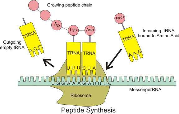
9. After transcription, the next stage is called translation. In this stage the mRNA moves to a ribosome in the cytoplasm. Transfer RNA (tRNA) molecules with complementary triplets of bases temporarily pair with base triplets on the mRNA, bringing appropriate amino acids. As two amino acids are held side by side, a peptide bond forms between them. The ribosome moves along the mRNA molecule, so that appropriate amino acids are gradually linked together, following the sequence laid down by the base sequence on the mRNA.

Multiple-choice Test 

1 What is found in both DNA and messenger RNA (mRNA)?

A deoxyribose
B double helix
C sugar–phosphate chain
D thymine

2 In DNA extracted from rat bone marrow, 29% of the bases were found to be adenine.

What was the proportion of cytosine?

A 58%
B 42%
C 29%
D 21%

3 The diagram shows part of a nucleic acid.


What is represented by X?

A a base pair
B a nucleotide
C a polynucleotide
D a purine

4 Which statement about base pairing is not correct?

A Adenine can pair with either thymine or uracil.
B Thymine pairs only with adenine.
C Cytosine makes two hydrogen bonds with guanine.
D Purine bases only pair with pyrimidine bases.

5 Which statements describe RNA?

1 composed of phosphate, deoxyribose, adenine, cytosine, guanine and thymine
2 backbone is a ribose–phosphate chain
3 each molecule consists of two chains
4 consists of a chain of nucleotides linked through phosphates and sugars

A 1, 2 and 3 only
B 1 and 2 only
C 2 and 3 only
D 2 and 4 only

6 The diagram shows part of a DNA molecule before replication.


Which diagram shows a daughter molecule?

7 A single base substitution in the gene coding for the β-globin polypeptide results in a change in the amino acid sequence.

Which statements describe what happens when haemoglobin containing polypeptides coded from the sickle cell allele, HbS, is not combined with oxygen?

1 The haemoglobin molecules are much less soluble.
2 The haemoglobin molecules form long fibres.
3 Red cells become distorted in shape.
4 Red cells become stuck in small capillaries.

A 1, 2, 3 and 4
B 1, 2 and 3 only
C 2 and 4 only
D 3 and 4 only

8 What is synthesised during transcription?

A DNA
B mRNA
C tRNA
D polypeptide

9 A mutation takes place in a DNA triplet coding for the amino acid tyrosine. The triplet ATA is changed to ATG.

The mRNA codons for tyrosine are UAU and UAC.

The mRNA codons signalling ‘stop’ are UAA, UAG and UGA.

What is the effect of the mutation?

A The mutated triplet codes for ‘stop’.
B The mutated triplet codes for a different amino acid.
C The mutated triplet is meaningless.
D The mutated triplet still codes for tyrosine.

10 In most organisms, the mRNA codons signalling ‘stop’ in translation are UAA, UAG and UGA. In the microorganism Methanosarcina barkeri, UAG codes for an amino acid.
Which tRNA carrying an amino acid will be found in M. barkeri but not in most organisms?


Answers for Multiple - choice Test

1  C
2  D
3  B
4  C
5  D
6  B
7  A
8  B
9  D
10 C

End-of-chapter questions

1. What can be found  in both  DNA and  messenger  RNA   (mRNA)?

A double helix  structure
B sugar-phosphate    chain
C  ribose
D thymine

2. Which statement about base pairing in  nucleic acids is not correct?

A Adenine can  pair  with  either   thymine  or uracil.
B Guanine  only  pairs  with  cytosine.
C  Thymine can  pair  with  either  adenine   or  uracil.
D Uracil only pairs  with  adenine.

3. How many different arrangements  of four bases into triplets can be made?

A 3+4
B 3 x 4
34
D 43

4. Look at the structures  of nucleotides in Figure  below:


Draw  a nucleotide that could be found:
a  in either DNA  or RNA
b only in DNA
c only in RNA.

5. Distinguish berween a nucleotide and  a nucleic acid.

6. Copy the drawing and annotate it to explain the replication of  DNA.


7. Use Appendix 1 to find the sequence of amino acids that is coded by the following length of messenger RNA (mRNA):

AUGUUUCUUGAUUAA

The table shows all the possible  triplets of bases in a DNA molecule and what each codes for. The three-letter  abbreviation for each amino acid is, in most cases, the first three letters of its full name -  see Appendix 2.

Appendix 1 
DNA triplet codes 


Appendix 2 
Amino acid R groups


8. The  table  shows  all the  messenger RNA (mRNA) codons for the amino acid leucine.
Copy the table and write in, for each codon, the transfer RNA (tRNA) anticodon that would bind with  it and the DNA triplet from which it was transcribed.
                            
mRNAcodon  
tRNA anticodon                     
DNA  triplet from which  mRNA was transcribed
UUA


UUG


CUU


CUC


CUA


CUG


9. In  most  people,  the  first  six amino  acids  in their β-globin  polypeptide chains are:
 
1    2     3     4     5    6

                                                              Val-His-Leu-Thr-Pro-Glu-rest of chain

The  DNA triplet coding for the sixth amino acid  (Glu) in most people is CTT. In some people this
DNA triplet is CAT.

a What type of mutation is the change from CTT to CAT?                                     [1]
b  Use Appendix 1 to identify the amino acid in the β-globin  polypeptide chains of people with  this  mutation.                                                                                                                  [1]
c  State the consequences for a person of having two copies of the mutated gene.  [1]
 [Total: 3]

10. Suggest why:

a  a mutation in which one nucleotide of a triplet code is altered often makes no difference to the protein molecule  coded by the DNA                                                                       [2]
b  the  addition or deletion of three nucleotides in the DNA sequence of a gene often has less effect  on  the encoded protein  than the addition or deletion of a single nucleotide.         [4]
 [Total: 3]

11. Copy and complete the following table to distinguish between the processes of transcription     and   translation.


Transcription
Translation
site in cell where process occurs


molecule  used as a template  in process


molecule  produced  by process


component  molecules  used in process


other molecules that are essential for the process to occur


12. The drawing shows polyribosomes.


a Name X, Y and Z.                                                                                                          [3]
b  In which direction are the ribosomes moving? Explain how you were able to decide on their direction of movement.                                                                                                     [2]
                                                                                                                                  [Total: 5]

                
Organism  (tissue)
Relative  numbers of bases
A
C
G
T
Ox (spleen)
27.9
20.8
22.7
27.3
Ox (thymus)
28.2
21.2
21.5
27.8
Yest
31.3
17.1
18.7
32.9
Virus with single-stranded DNA
24.3
18.2
24.5
32.3

Explainwhy:

a the relative numbers of each base in ox spleen and thymus are the same, within experimental error                                                                                                                                                      [2]
b the relative numbers of each base in yeast are different from those in ox spleen or thymus [2]
c  the relative number of the bases A and T, or of C and G, are similar in ox and yeast          [2]
d in the virus, the relative numbers of A and T, and  of C and G, are not similar.                   [2]

[Total: 8]

End-of-chapter answers

1 B
2 C
3 D

4


5 nucleotide:
 a molecule made up of a pentose sugar, a phosphate group and a nitrogenous base; 
 nucleic acid:
 a polymer of nucleotides/a polynucleotide;

6 Labels should include: 
 parent molecule;
 daughter molecules;
 parent/old strand acts as template;
 new strands made from nucleotides binding to old strands by complementary base pairing;
7 Met-Phe-Pro-Asp-[stop];
8

Exam-style questions

9  

9 a gene mutation/substitution; [1]
   b Val/valine;                         [1]
  c sickle cell anaemia;           [1]
                                       [Total: 3]

10 a many amino acids have more than one triplet code;  so sequence of amino acids is unchanged;                                                                                                                                                             [2]
    b adding or deleting three nucleotides may add or remove the coding for one amino acid;
this may not affect the final shape of the protein;
adding or deleting one nucleotide affects the arrangement of all subsequent triplets;
this ‘frameshift’ may alter the coding of all amino acids following the addition or deletion;
a triplet may be altered to a stop signal;                                                                                  [max. 4]

 [Total: 6]
11

Award 1 mark for each correct row. [5]

 12 a X mRNA;
        Y ribosome;
        Z (poly)peptide chain/chain of amino acids; [3]
    b from left to right;
       increasing length of polypeptide chain; [2]
 [Total: 5]

13 a the DNA in the spleen and thymus of the same organism is the same;
         the same genes are present in both organs; [2]
    b the DNA in different species is diff erent;
         different genes are present; [2]
   c  DNA has double helix/is double stranded;
     the numbers of A and T, and of C and G, are similar because A pairs with T and C pairs
     with G;                                 [2]
  d the DNA is single stranded;
         no base pairing occurs; [2]

 [Total: 8]


Gene mutation, sickle cell anaemia

A gene mutation is a change in the sequence of nucleotides that may result in an altered polypeptide.


A mutation is a random, unpredictable change in the DNA in a cell. It may be:

• a change in the sequence of bases in one part of a DNA molecule
• an addition of extra DNA to a chromosome or a loss of ONA from it
• a change in the total number of chromosomes in a cell.

Mutations are most likely to occur during DNA replication, for example when a 'wrong' base may slot into position in the new strand being built. Almost all of these mistakes are immediately repaired by enzymes, but some may persist.

Single point mutation. 
A change in the sequence of bases in DNA may result in a change in the sequence of amino acids in a protein. (Note that this does not always happen, because there is more than one triplet that codes for each amino acid, so a change in a triplet may not change the amino acid that is coded for.) This in turn may result in a change in the 3-D structure of the protein and therefore the way that it behaves.

Sickle cell anaemia

An example of a mutation is a change in the gene that codes for one of the polypeptides in a Hb molecule. In the genetic disease sickle cell anaemia, the gene that codes for the β polypeptide has the base T where it should have the base A. This means that one triplet is different, so a different amino acid is used when the polypeptide chain is constructed on a ribosome.


The abnormal β polypeptide has the amino acid valine where it should have the amino acid glutamic acid. The normal form of Hb is called HbA, the abnormal Hb is called sickle cell Hb (HbS)


These amino acids are on the outside of the Hb molecule when it takes up its tertiary and quaternary shapes. 
  • Glutamic acid is a hydrophilic amino acid. It interacts with water molecules, helping to make the haemoglobin molecule soluble. 
  • Valine is a hydrophobic amino acid. It does not interact with water molecules, making the haemoglobin molecule less soluble.

When the abnormal Hb is in an area of low oxygen concentration, the Hb molecules stick to one another, forming a big chain of molecules that is not soluble and therefore forms long fibres. This pulls the red blood cells out of shape, making them sickle-shaped instead of round. They are no longer able to move easily through the blood system and may get stuck in capillaries. This is very painful and can be fatal.


 Syllabus 2015

(d) describe the way in which the nucleotide sequence codes for the amino acid sequence in a polypeptide with reference to the nucleotide sequence for HbA (normal) and HbS (sickle cell) alleles of the gene for the β-globin polypeptide;


Syllabus 2016

6.2 Protein synthesis 

b) state that a gene mutation is a change in the sequence of nucleotides that may result in an altered polypeptide

c) describe the way in which the nucleotide sequence codes for the amino acid sequence in a polypeptide with reference to the nucleotide sequence for HbA (normal) and HbS (sickle cell)
alleles of the gene for the β-globin polypeptide


The genetic code - protein synthesis

The genetic code specifies the amino acids that are assembled to make polypeptides. The way that DNA codes for polypeptides is central to our understanding of how cells and organisms function.


A polypeptide is coded for by a gene and that a gene is a sequence of nucleotides that forms part of a DNA molecule.

The sequence of bases in a DNA molecule is a code that determines the sequence in which amino acids are linked together when making a protein molecule. A sequence of DNA nucleotldes that codes for 1 polypeptide, or for 1protein, is known as a gene.

The sequence of amino acids in a protein - its primary structure determines its 3-dimensional shape and therefore its properties and functions. For example, the primary structure of an enzyme determines the shape of its active site, and therefore the substrate with which it can bind.

A series of 3 bases in a DNA molecule, called a base triplet, codes for 1 amino acid. The DNA strand that is used in protein synthesis is called the template strand. For example, this is the sequence of amino acids coded for by the template strand of a particular length of DNA:


There are 20 amino acids. Because there are 4 bases, there are 43 - 64 different possible combinations of bases in a triplet. Some amino acids therefore are coded for by more than 1 triplet. For example, the triplets AAA and AAG both code for the amino acid phenylalanine. The code is therefore said to be degenerate.

Protein synthesis 

Proteins are made on the ribosomes in the cytoplasm, by linking together amino acids through peptide bonds. The sequence in which the amino acids are linked is determined by the sequence of bases on a length of DNA in the nucleus.


Translation and transcription


TRANSCRIPTION

The first step in protein synthesis is the transcription of messenger RNA (mRNA) from a DNA gene in the nucleus. 
  • In the nucleus, the double helix of the DNA is unzipped, exposing the bases on each strand.
  • There are 4 types of free RNA nucleotides in the nucleus, with the bases A, C,G and U. The RNA nucleotides form H bonds with the exposed bases on the template strand of the DNA. 

  • They pair up like this:


  • As the RNA nucleotides slot into place next to their complementary bases on the DNA, the enzyme RNA polymerase links them together (through their sugar and phosphate groups) to form a long chain of RNA nucleotides. This is an mRNA molecule. It contains a complementary copy of the base sequence on the template strand of part of a DNA molecule. 

TRANSLATION 
  • If the DNA coded for tRNA and rRNA, then the transcript is cut and the tRNA and rRNA are released.
  • If the DNA codes for protein, then the information carried in the sequence of RNA (mRNA) must be translated into a sequence of amino acids. Because we are changing “languages”, moving from a language of nucleotides to a language of amino acids, this process is called translation.
  • The process of translating information from RNA to protein will require:

                   - mRNA (copy of the original document)
                   - genetic code (translational dictionary)
                   - tRNA carrying the amino acid (a translator)
                   - ribosome (a writing desk, a place to do the work).
  • The mRNA molecule breaks away from the DNA, and moves out of the nucleus into the cytoplasm. In the cytoplasm, 20 different types of amino acids are present. 

  • There are also many different types of transfer RNA (tRNA) molecules. Each tRNA molecule is made up of a single strand of RNA nucleotides, twisted round on itself to form a clover-leaf shape. There is a group of 3 exposed bases, called an anticodon. There is also a position at which a particular amino acid can be loaded by a specific enzyme.

tRNA molecule. Credit Pearson. 


  • tRNA becomes attached to a ribosome. Triplets of nucleotides (codons) on the mRNA molecule fit into the anticodons of transfer RNA molecules that carry amino acids around inside the cell. 
  • The amino acid that can be loaded onto the tRNA is determined by the base sequence of its anticodon. For example, a tRNA whose anticodon is UAC will be loaded with the amino acid methionine.


    • A tRNA molecule with the complementary anticodon to the first codon on the mRNA, and carrying its appropriate amino acid, slots into place next to it in the ribosome, and hydrogen bonds form between the bases. Then a second tRNA does the same with the next mRNA codon.
    • Two codons fit into a groove in the ribosome. The first codon is generally AUG, which is known as a start codon. It codes for the amino acid methionine.
    • The amino acids carried by the two adjacent tRNAs are then linked by a peptide bond.
    • The mRNA is then moved along one place in the ribosome, and a third tRNA slots into place against the next mRNA codon. A third amino acid is added to the chain.


    Credit: Pearson.

    • The ribosomes combine the amino acids to form the finished protein.


      • This continues until a stop codon is reached on the mRNA. This is a codon that does not code for an amino acid, such as UGA. The polypeptide (long chain of amino acids) that has been formed breaks away.

      • This process of building a chain of amino acids following the code on an mRNA molecule is called translation.

       
       Syllabus 2015

      (c) state that a polypeptide is coded for by a gene and that a gene is a sequence of nucleotides that forms part of a DNA molecue and state that a mutation is a change in the sequence that may result in an altered polypeptide;

      (e) describe how the information on DNA is used during transcription and translation to construct polypeptides, including the role of messenger RNA (mRNA), transfer RNA (tRNA) and the ribosomes (for genetic dictionaries see section 5);


      Syllabus 2016 

      6.2 Protein synthesis 

      The genetic code specifies the amino acids that are assembled to make polypeptides. The way that
      DNA codes for polypeptides is central to our understanding of how cells and organisms function.

      a) state that a polypeptide is coded for by a gene and that a gene is a sequence of nucleotides that forms part of a DNA molecule

      d) describe how the information in DNA is used during transcription and translation to construct polypeptides, including the role of messenger RNA (mRNA), transfer RNA (tRNA) and the ribosomes

      Wednesday, 5 November 2014

      DNA structurer and replication

      DNA molecule consists of nucleotides in which the sugar component is deoxyribose whereas the RNA molecule has nucleotides in which the sugar is a ribose.


      Nucleotides

      Roles:
      • are monomers for nucleic acid polymers, such as DNA and RNA. The genetic material (DNA) is a polymer of 4 different nucleotides. The genetic information is coded in the sequence of nucleotides in a DNA molecule. 
      • adenosine triphosphate (ATP) - the nucleotide molecule that doesn't include the phosphate group - is energy carrier in metabolic pathways. 
      • are components of some important coenzymes:  flavin adenine dinucleotide (FAD), nicotinamide adenine dinucleotide phosphate (NADP) and Coenzyme A. 
      They consist of:
      1. A 5-carbon sugar (deoxyribose in DNA; ribose in RNA)
      2. a phosphate group. 
      3. a 1 or 2 ring nitrogen-containing base

      The bases are usually referred to by their first letters:
      • A = adenin, 
      • G =guamine, 
      • C = cystosine, 
      • T = thymine (methyl +uracil)
      • U = uracil.
      • A and G = purine bases (2 carbon-nitrogen rings). 
      • C, T and U = pyrimidine bases (1 carbon-nitrogen ring).


      Nucleotides bond together to make nucleic acids. They form covalent bonds between the phosphate group of one and the sugar of another. This takes place through a condensation reaction.


      Structure of DNA and RNA

      DNA and RNA are polynucleotides (long chains of nucleotides).
      DNA = A, G, C, T
      RNA = A, G, C, U


      + RNA molecule -  a single strand (may be folded up on itself). 


      + DNA molecule 
      • 2 strands run in opposite directions (anti-parallel) and twist round each other --> double helix. 
      • There are H bonds between the bases on the 2 strands. 
      • H bonding only occurs between A-T and C-G (complementary base pairing).  

      The significance of complementary base pairing
      • Only certain pairings of bases are possible ( A-T and G-C) => 2 strands of the double helix are complementary, each the predictable counterpart of the other.
      • Since the 2 strands of DNA are complimentary, they can separate from one another and each can serve as a template for building a new partner (if you know the sequence of one DNA strand then you can easily figure out the sequence of the other strand).
      • Thus, DNA replication is semi-conservative, with each of the two daughter DNA molecules having one old strand derived from the parent and one newly made strand.
      • The complementary base pairing results in the two daughter DNA molecules being identical.

      DNA Replication

      New DNA molecules need to be made before a cell can divide. The 2 daughter cells must each receive a complete set of DNA. The base sequences on the new DNA molecules must be identical with those on the original set. DNA replication takes place in the nucleus, during interphase.

      Semi-conservative DNA replication

      • Hydrogen bonds between the bases along part of the two strands are broken. This 'unzips' part of the molecule, separating the two strands.


      • Nucleotides that are present in solution in the nucleus are moving randomly around. By chance, a free nucleotide will bump into a newly exposed one with which it can form hydrogen bonds. Free nucleotides therefore pair up with the nucleotides on each of the DNA strands, always A with T and C with G. DNA polymerase links together the phosphate and deoxyribose groups of adjacent
      nucleotides.


      • As each strand retains half of the original DNA material, this method of replication is called semi-consservative. 


      3 types of DNA replication 
      • Semi-conservative replication: each new DNA has 1 old strand and 1 new one. 
      • Conservative replication: each new DNA has 2 new strands containing all of the new DNA base pairs. The two original template DNA strands stay together in a double helix. 
      • Dispersive replication: each new DNA has distinct regions of DNA composed of either both original strands or both new strands.


       Syllabus 2015

       (a) describe the structure of RNA and DNA and explain the importance of base pairing and the different hydrogen bonding between bases (includes reference to adenine and guanine as purines and to cytosine, thymine and uracil as pyrimidines. Structural formulae for bases is not required but the  recognition that purines have a double ring structure and pyrimidines have a single ring structure should be included);

      (b) explain how DNA replicates semi-conservatively during interphase;


      Syllabus 2016


      6.1 Structure and replication of DNA


      Understanding the structure of nucleic acids allows an understanding of their role in the storage of genetic information and how that information is used in the synthesis of proteins.

      a) describe the structure of nucleotides, including the phosphorylated nucleotide ATP (structural formulae are not required)

      b) describe the structure of RNA and DNA and explain the importance of base pairing and the different hydrogen bonding between bases (include reference to adenine and guanine as purines and to cytosine, thymine and uracil as pyrimidines. Structural formulae for bases are not required but the recognition that purines have a double ring structure and pyrimidines have a single ring structure should be included)

      c) describe the semi-conservative replication of DNA during interphase